Yonsei Med J.
2013 Jul;54(4):907-911. 10.3349/ymj.2013.54.4.907.
Robotic Mechanical Localization of Prostate Cancer Correlates with Magnetic Resonance Imaging Scans
- Affiliations
-
- 1Department of Urology, Urological Science Institute, Yonsei University College of Medicine, Seoul, Korea. khrha@yuhs.ac
- 2School of Mechanical, Aerospace & Systems Engineering, Department of Mechanical Engineering, Korea Advanced Institute of Science and Technology, Daejeon, Korea.
- KMID: 2158225
- DOI: http://doi.org/10.3349/ymj.2013.54.4.907
Abstract
- PURPOSE
To evaluate the concordance of cancer location of the tissue mapping from a mechanical pressure transducer with magnetic resonance imaging (MRI) scans.
MATERIALS AND METHODS
A total of 60 indentations were performed on 5 prostate specimens obtained after radical prostatectomy utilizing a robotic indentation system. The mechanical elastic moduli of suspected malignant lesions were calculated and mapped, and their locations were compared with suspicious areas of malignancy on MRI scans.
RESULTS
The concordance rate between the location mapping from the robotic indentation system and MRI scans results was 90.0% (54/60). The sensitivity and specificity of the robotic indentation system were 87.9% (29/33) and 92.6% (25/27), respectively. The positive predictive value and negative predictive value were 93.5% (29/31) and 93.1% (27/29), respectively.
CONCLUSION
The locations of malignant lesions derived from our robotic indentation system correlated strongly with the locations of suspected areas of malignancy on MRI scans. Our robotic system may provide a more targeted biopsy of the prostate than conventional non-targeted systemic biopsy, possibly improving the diagnostic accuracy of prostatic biopsies for cancer.
Keyword
MeSH Terms
Figure
-
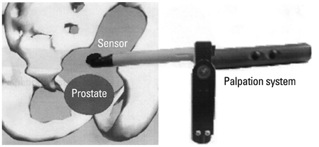
Fig. 1 Schematics of experimental setup.
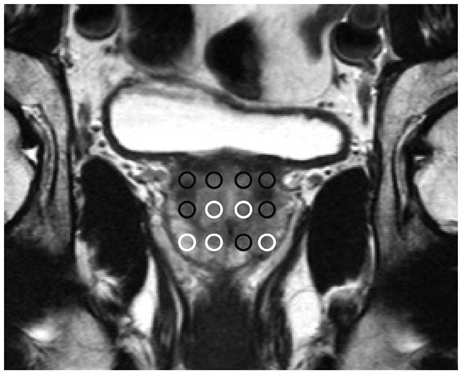
Fig. 2 The Example of localization process of lesions suspicious of cancer on MRI scans under normal property criteria of the prostate tissue (black circle: suspicious lesion, white circle: not suspicious lesion). MRI, magnetic resonance imaging.
Reference
-
1. Siegel R, Ward E, Brawley O, Jemal A. Cancer statistics, 2011: the impact of eliminating socioeconomic and racial disparities on premature cancer deaths. CA Cancer J Clin. 2011; 61:212–236.2. Borden LS Jr, Wright JL, Kim J, Latchamsetty K, Porter CR. An abnormal digital rectal examination is an independent predictor of Gleason > or =7 prostate cancer in men undergoing initial prostate biopsy: a prospective study of 790 men. BJU Int. 2007; 99:559–563.
Article3. Gosselaar C, Roobol MJ, van den Bergh RC, Wolters T, Schröder FH. Digital rectal examination and the diagnosis of prostate cancer--a study based on 8 years and three screenings within the European Randomized Study of Screening for Prostate Cancer (ERSPC), Rotterdam. Eur Urol. 2009; 55:139–146.
Article4. Yossepowitch O. Digital rectal examination remains an important screening tool for prostate cancer. Eur Urol. 2008; 54:483–484.
Article5. Mettlin C, Lee F, Drago J, Murphy GP. The American Cancer Society National Prostate Cancer Detection Project. Findings on the detection of early prostate cancer in 2425 men. Cancer. 1991; 67:2949–2958.
Article6. Bozeman CB, Carver BS, Caldito G, Venable DD, Eastham JA. Prostate cancer in patients with an abnormal digital rectal examination and serum prostate-specific antigen less than 4.0 ng/mL. Urology. 2005; 66:803–807.
Article7. Park KK, Chung MS, Chung SY, Kim JH, Chung BH. Effects of post biopsy digital rectal compression on improving prostate cancer staging using magnetic resonance imaging in localized prostate cancer. Yonsei Med J. 2013; 54:81–86.
Article8. Ahn B, Lorenzo EI, Rha KH, Kim HJ, Kim J. Robotic palpation-based mechanical property mapping for diagnosis of prostate cancer. J Endourol. 2011; 25:851–857.
Article9. Remzi M, Fong YK, Dobrovits M, Anagnostou T, Seitz C, Waldert M, et al. The Vienna nomogram: validation of a novel biopsy strategy defining the optimal number of cores based on patient age and total prostate volume. J Urol. 2005; 174(4 Pt 1):1256–1260.
Article10. Djavan B, Ravery V, Zlotta A, Dobronski P, Dobrovits M, Fakhari M, et al. Prospective evaluation of prostate cancer detected on biopsies 1, 2, 3 and 4: when should we stop? J Urol. 2001; 166:1679–1683.
Article11. Rosen J, Brown JD, De S, Sinanan M, Hannaford B. Biomechanical properties of abdominal organs in vivo and postmortem under compression loads. J Biomech Eng. 2008; 130:021020.
Article12. Samur E, Sedef M, Basdogan C, Avtan L, Duzgun O. A robotic indenter for minimally invasive measurement and characterization of soft tissue response. Med Image Anal. 2007; 11:361–373.
Article13. Nava A, Mazza E, Furrer M, Villiger P, Reinhart WH. In vivo mechanical characterization of human liver. Med Image Anal. 2008; 12:203–216.
Article14. Scattoni V, Zlotta A, Montironi R, Schulman C, Rigatti P, Montorsi F. Extended and saturation prostatic biopsy in the diagnosis and characterisation of prostate cancer: a critical analysis of the literature. Eur Urol. 2007; 52:1309–1322.
Article15. Bonekamp D, Jacobs MA, El-Khouli R, Stoianovici D, Macura KJ. Advancements in MR imaging of the prostate: from diagnosis to interventions. Radiographics. 2011; 31:677–703.
Article16. Lattouf JB, Grubb RL 3rd, Lee SJ, Bjurlin MA, Albert P, Singh AK, et al. Magnetic resonance imaging-directed transrectal ultrasonography-guided biopsies in patients at risk of prostate cancer. BJU Int. 2007; 99:1041–1046.
Article17. Stoianovici D, Song D, Petrisor D, Ursu D, Mazilu D, Muntener M, et al. "MRI Stealth" robot for prostate interventions. Minim Invasive Ther Allied Technol. 2007; 16:241–248.
Article
- Full Text Links
-
- Actions
-
Cited
- CITED
-
- Close
- Share
-
- Similar articles
-
- Medical imaging of prostate cancer
- Multiparametric MRI in the Detection of Clinically Significant Prostate Cancer
- Magnetic resonance imaging-transrectal ultrasound fusion image-guided prostate biopsy: Current status of the cancer detection and the prospects of tailor-made medicine of the prostate cancer
- The Use of Magnetic Resonance Imaging in the Prostate Cancer Primary Diagnostic Pathway: Is It Ready for Primetime?
- Added values of transrectal ultrasonography to magnetic resonance imaging in characterizing prostate cancer: A narrative review
