Dysplastic Nevus with Eruptive Melanocytic Lesions That Developed during Nilotinib Therapy for Chronic Myeloid Leukemia
- Affiliations
-
- 1Department of Dermatology, The Catholic University of Korea, College of Medicine, Seoul, Korea. hjpark@catholic.ac.kr
- KMID: 2157466
- DOI: http://doi.org/10.5021/ad.2015.27.6.782
Abstract
- No abstract available.
Figure
-
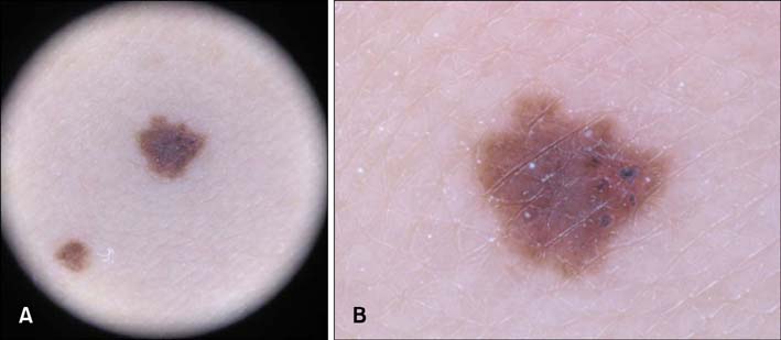
Fig. 1 Dermoscopic picture showing structureless pigmentation with asymmetric and irregular and fuzzy borders, and several blackish globules.
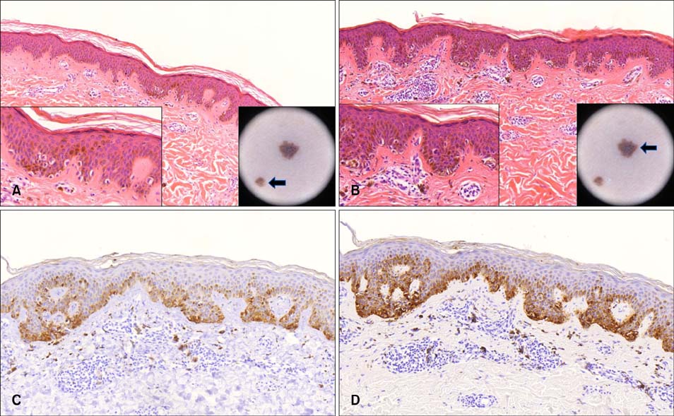
Fig. 2 (A) Histopathological picture of the benign melanocytic lesion showing slightly elongated, clubbed rete ridges and intermittently distributed single melanocytes at the tip and side of the rete ridges. Lesional melanocytes did not show dysplasia (H&E, ×100; left inset: ×400; right inset: clinical picture). (B) Histopathological picture of the atypical pigmented lesion showing elongated, fused rete ridges and contiguously distributed single melanocytes and a few small nests at the dermal-epidermal junction. In addition, eosinophilic fibroplasias and mild perivascular infiltrate were observed. Lesional melanocytes were observed to have moderately enlarged nuclei compared with most normal melanocytes and slightly irregular, hyperchromatic nuclei on the high-power field (×100; left inset: ×400; right inset: clinical picture). (C) Melan-A immunoreactivity of contiguously distributed melanocytes (×100). (D) HMB45 immunoreactivity of contiguously distributed melanocytes (×100).
Reference
-
1. Bovenschen HJ, Tjioe M, Vermaat H, de Hoop D, Witteman BM, Janssens RW, et al. Induction of eruptive benign melanocytic naevi by immune suppressive agents, including biologicals. Br J Dermatol. 2006; 154:880–884.
Article2. Zattra E, Fortina AB, Bordignon M, Piaserico S, Alaibac M. Immunosuppression and melanocyte proliferation. Melanoma Res. 2009; 19:63–68.
Article3. Kong HH, Sibaud V, Chanco Turner ML, Fojo T, Hornyak TJ, Chevreau C. Sorafenib-induced eruptive melanocytic lesions. Arch Dermatol. 2008; 144:820–822.
Article4. Reutter JC, Long EM, Morrell DS, Thomas NE, Groben PA. Eruptive post-chemotherapy in situ melanomas and dysplastic nevi. Pediatr Dermatol. 2007; 24:135–137.
Article5. Tran A, Tawbi HA. A potential role for nilotinib in KIT-mutated melanoma. Expert Opin Investig Drugs. 2012; 21:861–869.
- Full Text Links
-
- Actions
-
Cited
- CITED
-
- Close
- Share
-
- Similar articles
-
- Eruptive Melanocytic Nevi Induced by Radotinib
- A Case of Congenital Melanocytic Nevus Combined with an Epidermal Cyst
- Nilotinib-Induced Acute Interstitial Nephritis during the Treatment of Chronic Myeloid Leukemia
- A Case of Dysplastic Melanocytic Nevus
- A Case of a Compound Nevus That Developed as Papillomatous Melanocytic Nevus
