Ann Dermatol.
2011 Oct;23(Suppl 2):S226-S230. 10.5021/ad.2011.23.S2.S226.
Acantholytic Anaplastic Extramammary Paget's Disease: A Case Report and Review of the Literature
- Affiliations
-
- 1Department of Dermatology, College of Medicine, Kyung Hee University, Seoul, Korea. bellotte@hanmail.net
- KMID: 2156796
- DOI: http://doi.org/10.5021/ad.2011.23.S2.S226
Abstract
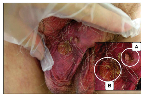
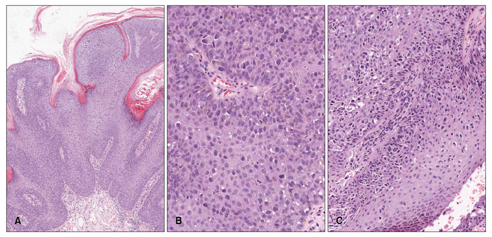
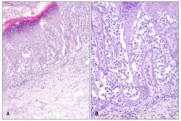
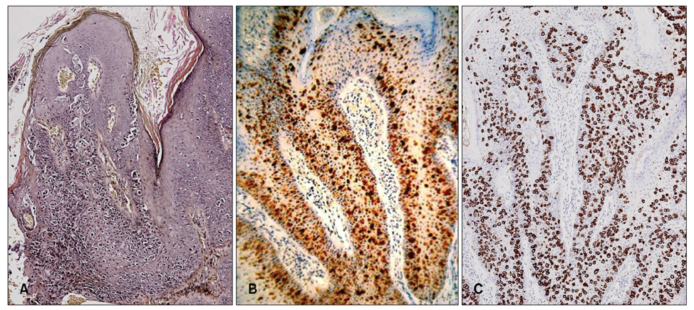
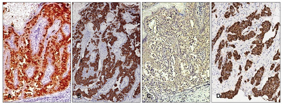
- Extramammary Paget's disease (EMPD) is an uncommon intraepithelial neoplasm that most commonly arises on the vulva and perianal region. Very few cases of EMPD revealing a histological Bowenoid appearance have been reported. This study describes scrotal EMPD presenting with histological features of Bowen's disease in a 79-year-old man. He presented with a 5-year history of a pruritic erythematous plaque and a verrucous papule on the scrotum. The verrucous papule histopathologically showed Bowenoid features, and the erythematous plaque demonstrated acantholytic EMPD. Immunohistochemical findings revealed strong expression for carcinoembryonic antigen, Cam 5.2, epithelial membrane antigen, cytokeratin (CK) 7, and pancytokeratin (AE1/AE3) in both areas, but negative CK20 staining, supporting the overall diagnosis of primary acantholytic anaplastic EMPD. This is the first reported case of acantholytic anaplastic EMPD in the Korean literature.
MeSH Terms
Figure
Reference
-
1. Williamson JD, Colome MI, Sahin A, Ayala AG, Medeiros LJ. Pagetoid bowen disease: a report of 2 cases that express cytokeratin 7. Arch Pathol Lab Med. 2000. 124:427–430.2. Quinn AM, Sienko A, Basrawala Z, Campbell SC. Extramammary Paget disease of the scrotum with features of Bowen disease. Arch Pathol Lab Med. 2004. 128:84–86.
Article3. Cannavò SP, Guarneri F, Napoli P. Extramammary Paget's disease of the scrotum with Bowenoid features. Eur J Dermatol. 2006. 16:203–204.4. Rayne SC, Santa Cruz DJ. Anaplastic Paget's disease. Am J Surg Pathol. 1992. 16:1085–1091.
Article5. Mobini N. Acantholytic anaplastic Pagets disease. J Cutan Pathol. 2009. 36:374–380.
Article6. Mazoujian G, Pinkus GS, Haagensen DE Jr. Extramammary Paget's disease--evidence for an apocrine origin. An immunoperoxidase study of gross cystic disease fluid protein-15, carcinoembryonic antigen, and keratin proteins. Am J Surg Pathol. 1984. 8:43–50.7. Hawley IC, Husain F, Pryse-Davies J. Extramammary Paget's disease of the vulva with dermal invasion and vulval intra-epithelial neoplasia. Histopathology. 1991. 18:374–376.
Article8. Orlandi A, Piccione E, Sesti F, Spagnoli LG. Extramammary Paget's disease associated with intraepithelial neoplasia of the vulva. J Eur Acad Dermatol Venereol. 1999. 12:183–185.
Article9. Matsumoto M, Ishiguro M, Ikeno F, Ikeda M, Kamijima R, Hirata Y, et al. Combined Bowen disease and extramammary Paget disease. J Cutan Pathol. 2007. 34:Suppl 1. 47–51.
Article

