Ann Dermatol.
2011 Oct;23(Suppl 2):S208-S210. 10.5021/ad.2011.23.S2.S208.
Acral-type Malignant Acanthosis Nigricans Associated with Gastric Adenocarcinoma
- Affiliations
-
- 1Department of Dermatology, School of Medicine, Chungnam National University, Daejeon, Korea. jhoon@cnu.ac.kr
- KMID: 2156791
- DOI: http://doi.org/10.5021/ad.2011.23.S2.S208
Abstract
- Acanthosis nigricans is a symmetric eruption characterized by the presence of a hyperpigmented, velvety cutaneous thickening, that can develop on any part of the body, but characteristically affects the flexural areas of the body. The velvety hyperkeratotic lesions can be located on the dorsum of the hands and feet in dark-skinned people in the form of a variant of acanthosis nigricans called as acral acanthotic anomaly or acral type acanthosis nigricans. Although acanthosis nigricans is associated with malignant tumors, particularly gastric carcinoma, acral type acanthosis nigricans has never been reported to be associated with gastric adenocarcinoma. In our present study, we describe a case of 58-year-old man with acral type acanthosis nigricans and its association with carcinoma of the stomach; a marked improvement was seen in the skin condition of the patient with chemotherapy.
Figure
-

Fig. 1 Velvety, ill-defined hyperpigmentation and papillary hypertrophy are seen at the joints of the fingers (A) and toes (B).
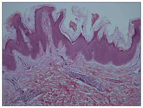
Fig. 2 The lesion at the finger joint shows hyperkeratosis, papillomatosis, and hyperpigmentation of the basal layer. Some dermal papillae were projected upward in the form of finger-like projections (H&E, ×100).
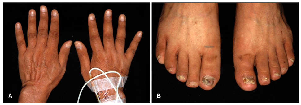
Fig. 3 (A, B) Treatment with anti-cancer chemotherapy improved the skin symptoms of acanthosis nigricans.
Cited by 1 articles
-
Clinical and Histopathologic Study of Confluent and Reticulated Papillomatosis by Anatomic Site and Age
Young Min Cho, Kyung Eun Jung, Dae Won Koo, Joong Sun Lee
Ann Dermatol. 2018;30(5):550-555. doi: 10.5021/ad.2018.30.5.550.
Reference
-
1. Christine AD, Lucinda SB, Stephen PS. Wolff K, Goldsmith LA, Katz SI, Glichrest BA, Paller AS, Leffel DJ, editors. Cutaneous manifestation of internal malignant disease. Fitzpatrick's dermatology in general medicine. 2008. 7th ed. New York: McGraw-Hill;1494–1498.2. Curth HO. Classification of acanthosis nigricans. Int J Dermatol. 1976. 15:592–593.
Article3. Schwartz RA. Acral acanthotic anomaly (AAA). J Am Acad Dermatol. 1981. 5:345–346.
Article4. Melczer N, Dvorszky C. Acanthosisnigricans bei dermatofibroma protuberans mit multiplen hautmetastasen. Hautarzt. 1957. 8:54–58.5. Song JY, Lim JH, Kim CW, Kim HO. A case of acral type acanthosis nigricans associated with lymphoma. Korean J Dermatol. 2002. 40:841–843.6. Koyama S, Ikeda K, Sato M, Shibahara K, Yuhara K, Fukutomi H, et al. Transforming growth factor-alpha (TGF-α)-producing gastric carcinoma with acanthosis nigricans: an endocrine effect of TGF-α in the pathogenesis of cutaneous paraneoplastic syndrome and epithelial hyperplasia of the esophagus. J Gastroenterol. 1997. 32:71–77.
Article7. Ellis DL, Kafka SP, Chow JC, Nanney LB, Inman WH, McCadden ME, et al. Melanoma, growth factors, acanthosis nigricans, the sign of Leser-Trelat and multiple acrochordons. N Engl J Med. 1987. 317:1582–1587.
Article
- Full Text Links
-
- Actions
-
Cited
- CITED
-
- Close
- Share
-
- Similar articles
-
- A Case of Acral Type Acanthosis Nigricans Associated with Lymphoma
- A Case of Malignant Acanthosis Nigricans Associated with Gastric Adenocarcinoma
- Malignant Acanthosis Nigricans Associated with Stomach Adenocarcinoma
- A case of malignant acanthosis nigricans associated with gastric adenocarcinoma
- Malignanr Acanthosis Nigricans: Report of a Case
