Ann Dermatol.
2011 Sep;23(Suppl 1):S123-S126. 10.5021/ad.2011.23.S1.S123.
Application of Sentinel Lymph Node Biopsy in Cutaneous Basosquamous Carcinoma
- Affiliations
-
- 1Clinic of Plastic and Reconstructive Surgery, Clinical Center Nis, 18000 Nis, Serbia. jankovic@jotel.co.rs
- 2Department of Pathology and Pathophysiology, Faculty of Cosmetology and Aesthetics, Banja Luka, University Sinergy, Bjeljina, Bosnia and Herzegovina.
- 3Clinic of Dermatovenerology, Clinical Center Nis, 18000 Nis, Serbia.
- 4Institute of Pathology, Clinical Center Nis, 18000 Nis, Serbia.
- KMID: 2156769
- DOI: http://doi.org/10.5021/ad.2011.23.S1.S123
Abstract
- Basosquamous carcinoma of the skin is a relatively rare cutaneous neoplasm that has significant metastatic potential and a metastatic rate greater than that of basal cell and squamous cell carcinoma. We describe the use of lymphatic mapping and sentinel lymph node biopsy in a 63-year-old man after identification of basosquamous carcinoma. Sentinel lymph node biopsy, which is a standard tool to detect regional lymphatic metastasis in cutaneous melanoma, has been rarely employed to detect lymphatic metastasis of basosquamous carcinoma. The approach was successful in detecting a regional lymphatic metastasis of two nodal basins with minor morbidity. Sentinel lymph node biopsy may be useful for certain high-risk lesions of basosquamous carcinoma.
MeSH Terms
Figure
-

Fig. 1 Photograph of the lesion.
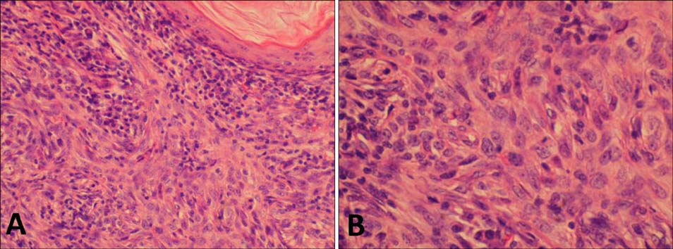
Fig. 2 Histopathological examination of the primary tumor. (A) Conventional basaloid tumor islands were admixed with more atypical tumor cells demonstrating higher grade cytologic atypia with squamous differentiation (H&E stain, ×200). (B) A high power view revealing BCC cells with squamous differentiation (H&E stain, ×400).

Fig. 3 SLN of the left axilla with metastatic BSC (CK AE1/AE3, ×10).
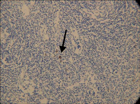
Fig. 4 SLN of the right axilla with isolated tumor cells of SCC(CK AE1/AE3, ×20).
Reference
-
1. Lee SJ, Lim HJ, Kim HY, Song CH, Kim BS, Lee WJ, et al. The feasibility of sentinel lymph node biopsy with a multidisciplinary cooperative team approach for the management of koreans with cutaneous malignant melanoma. Ann Dermatol. 2010. 22:26–34.
Article2. Ross AS, Schmults CD. Sentinel lymph node biopsy in cutaneous squamous cell carcinoma: a systematic review of the English literature. Dermatol Surg. 2006. 32:1309–1321.
Article3. Anadolu-Brasie R, Patel AR, Patel SS, Singh A, Nouri K. Nouri K, editor. Squamous Cell Carcinoma of the Skin. Skin cancer. 2008. New York: McGraw-Hill;104.
Article4. Martin RC 2nd, Edwards MJ, Cawte TG, Sewell CL, McMasters KM. Basosquamous carcinoma: analysis of prognostic factors influencing recurrence. Cancer. 2000. 88:1365–1369.
Article5. Bowman PH, Ratz JL, Knoepp TG, Barnes CJ, Finley EM. Basosquamous carcinoma. Dermatol Surg. 2003. 29:830–833.
Article6. Costantino D, Lowe L, Brown DL. Basosquamous carcinoma-an under-recognized, high-risk cutaneous neoplasm: case study and review of the literature. J Plast Reconstr Aesthet Surg. 2006. 59:424–428.7. Alam M, Ratner D. Cutaneous squamous-cell carcinoma. N Engl J Med. 2001. 344:975–983.8. von Domarus H, Stevens PJ. Metastatic basal cell carcinoma. Report of five cases and review of 170 cases in the literature. J Am Acad Dermatol. 1984. 10:1043–1060.
Article9. Doubrovsky A, De Wilt JH, Scolyer RA, McCarthy WH, Thompson JF. Sentinel node biopsy provides more accurate staging than elective lymph node dissection in patients with cutaneous melanoma. Ann Surg Oncol. 2004. 11:829–836.
Article
- Full Text Links
-
- Actions
-
Cited
- CITED
-
- Close
- Share
-
- Similar articles
-
- Sentinel Lymph Node Biopsy in the Oral Cavity Cancer
- The Number of Removed Lymph Nodes for an Acceptable False Negative Rate in Sentinel Lymph Node Biopsy for Breast Cancer
- Clinical Significance of Sentinel Lymph Node Detection in Vulvar Cancer
- Treatment of Malignant Melanoma Using Sentinel Lymph Node Dissection
- Validation and Controversy of Sentinel Node Biopsy for Breast Cancer
