Ann Dermatol.
2011 Sep;23(Suppl 1):S72-S74. 10.5021/ad.2011.23.S1.S72.
Development of Dermatomyofibroma in a Male Infant
- Affiliations
-
- 1Department of Dermatology, Ajou University School of Medicine, Suwon, Korea. maychan@ajou.ac.kr
- 2Department of Pathology, St Mary's/Duluth Clinic Health System, Duluth, MN, USA.
- KMID: 2156756
- DOI: http://doi.org/10.5021/ad.2011.23.S1.S72
Abstract
- Dermatomyofibroma is a rare benign cutaneous mesenchymal neoplasm of the fibroblasts and myofibroblasts. The majority of dermatomyofibromas present as red-brown discolored plaques or nodules, commonly located on the shoulder, upper arm, axilla, neck, and/or upper trunk. These lesions develop most frequently in young female patients at a mean of 28-years-of-age. Herein, a case of dermatomyofibroma is reported that developed in an infant. A 4-month-old boy presented with an ill-defined bluish firm plaque on the trunk that developed 1 month after birth. Histopathologically, there was proliferation of bland-looking spindle cells with fascicular arrangement in the dermis and subcutaneous tissue. Immunohistochemistry showed that most of the tumor cells expressed diffuse positivity for vimentin and smooth muscle actin, but were negative for S-100 protein, desmin, and CD34.
Keyword
MeSH Terms
Figure
-
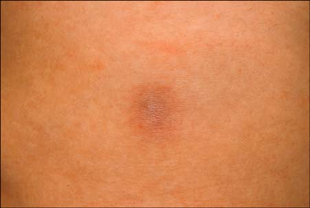
Fig. 1 An ill-defined bluish firm plaque on the left flank.
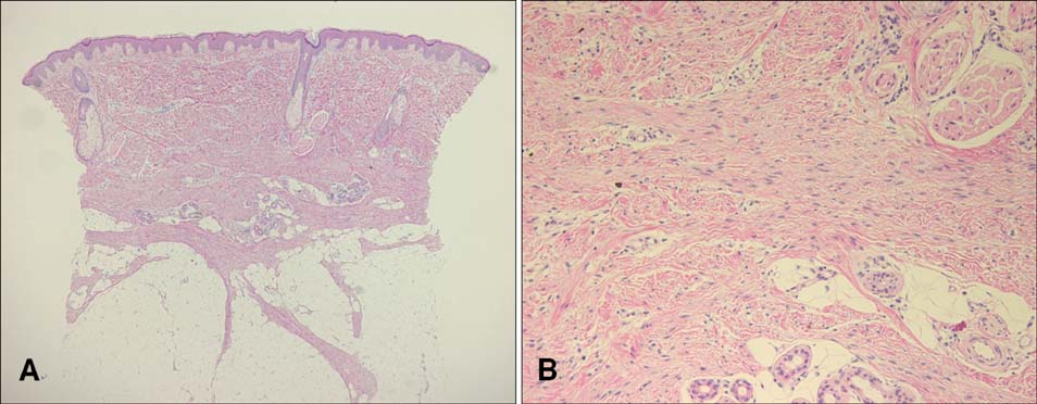
Fig. 2 Proliferation of bland-looking spindle cells with fascicular arrangement in the reticular dermis (A), extending focally into the subcutaneous tissue (B). There was no cytological atypia or mitotic figures (H&E; original magnification, A: ×40; B: ×200).
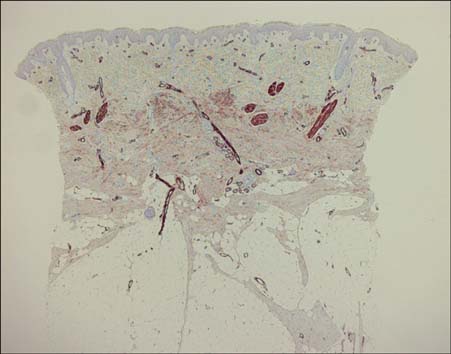
Fig. 3 Immunohistochemistry showed that most of the tumor cells expressed a diffuse positivity for smooth muscle actin (Original magnification, ×40).
Reference
-
1. Gilaberte Y, Coscojuela C, Doste D, Vera J, Requena L. Dermatomyofibroma in a male child. J Eur Acad Dermatol Venereol. 2005. 19:257–259.2. Mentzel T, Kutzner H. Dermatomyofibroma: clinicopathologic and immunohistochemical analysis of 56 cases and reappraisal of a rare and distinct cutaneous neoplasm. Am J Dermatopathol. 2009. 31:44–49.3. Kim SH, Oh JJ, Lee JH, Lee ES. A case of dermatomyofibroma in a 2 year old boy. Ann Dermatol. 2003. 15:68–70.
Article4. Mortimore RJ, Whitehead KJ. Dermatomyofibroma: a report of two cases, one occurring in a child. Australas J Dermatol. 2001. 42:22–25.
Article5. Rose C, Bröcker EB. Dermatomyofibroma: case report and review. Pediatr Dermatol. 1999. 16:456–459.
Article6. Kamino H, Reddy VB, Gero M, Greco MA. Dermatomyofibroma. A benign cutaneous, plaque-like proliferation of fibroblasts and myofibroblasts in young adults. J Cutan Pathol. 1992. 19:85–93.
Article7. Viglizzo G, Occella C, Calonje E, Nozza P, Rongioletti F. A unique case of multiple dermatomyofibromas. Clin Exp Dermatol. 2008. 33:622–624.
Article8. Prieto VG, Reed JA, Shea CR. Immunohistochemistry of dermatofibromas and benign fibrous histiocytomas. J Cutan Pathol. 1995. 22:336–341.
Article
