A Case of Fibroelastolytic Papulosis on the Neck of a Young Man
- Affiliations
-
- 1Department of Dermatology, Konkuk University School of Medicine, Seoul, Korea. 20050078@kuh.ac.kr
- 2Department of Pathology, ChungAng University School of Medicine, ChungAng University Medical Center, Seoul, Korea.
- KMID: 2156661
- DOI: http://doi.org/10.5021/ad.2011.23.2.193
Abstract
- Fibroelastolytic papulosis of the neck (FEPN) encompasses a spectrum of two disorders that were previously reported as pseudoxanthoma elasticum-like papillary dermal elastolysis (PXE-PDE) and white fibrous papulosis of the neck (WFPN). The clinical presentation of FEPN is asymptomatic to mildly pruritic whitish-yellow papules that may coalesce into cobblestone patterned plaques that resemble pseudoxanthoma elasticum (PXE). The histology is characterized by a decrease or loss of elastic fibers in the papillary dermis and this is sometimes accompanied by a minimal or nodular increase of dermal collagen fibers. We report here on a 28-year-old Korean man with asymptomatic, multiple, skin-colored to slightly yellowish, match-head sized, cobblestone-patterned papules on the neck, and these were histologically consistent with FEPN and the papules showed slightly increased dermal collagen associated with decreased and fragmented elastic fibers, elastin and tropoelastin. The pathogenesis of FEPN in this case might have been related with mild dermal inflammation, followed by fragmentation, elastolysis and increased dermal collagen.
Keyword
MeSH Terms
Figure
-

Fig. 1 Asymptomatic, multiple, yellowish to skin colored, match-head sized, cobblestone-patterned papules on the neck of a 28-year-old man. The lesions were more prominently observed when the head was rotated.
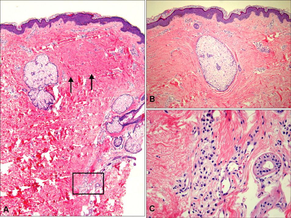
Fig. 2 (A) Histology of the papular skin lesion showed nodular increased dermal collagen in the upper dermis (arrow) with a focal inflammatory reaction (rectangle) in the lower dermis (H&E, ×40). (B) Dermal sclerosis due to the increased collagen around the sebaceous gland in the mid dermis (H&E, ×100). (C) The high power view of a rectangular area in (A) showed a few lymphocytes and plasma cells in the periadnexa (H&E, ×200).
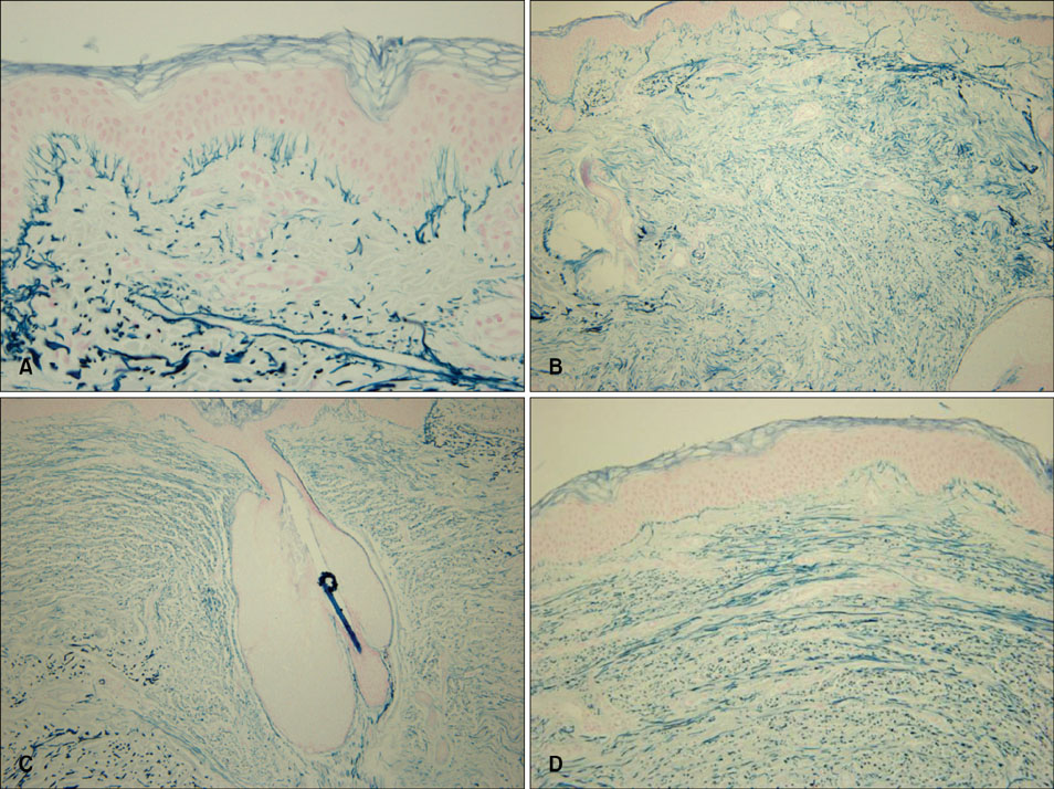
Fig. 3 (A) Verhoeff elastic stain shows decreased elastic fibers in the upper and mid dermal areas (Verhoeff elastic stain, ×100). (B) Victoria blue elastic stain shows decreased and fragmented elastic fibers in the dermis (Victoria blue elastic stain, ×200). (C) Immunohistochemical staining for the elastic fibers shows decreased and fragmented elastic fibers in the dermis (Immunoperoxidase staining with anti-elastic fiber antibody, ×200). (D) Immunohistochemical staining for tropoelastin shows these are decreased and fragmented in the dermis (Immunoperoxidase stain with anti-tropoelastin antibody, ×200).
Reference
-
1. Balus L, Amantea A, Donati P, Fazio M, Giuliano MC, Bellocci M. Fibroelastolytic papulosis of the neck: a report of 20 cases. Br J Dermatol. 1997. 137:461–466.
Article2. Rongioletti F, Rebora A. Pseudoxanthoma elasticum-like papillary dermal elastolysis. J Am Acad Dermatol. 1992. 26:648–650.
Article3. Shimizu H, Kimura S, Harada T, Nishikawa T. White fibrous papulosis of the neck: a new clinicopathologic entity? J Am Acad Dermatol. 1989. 20:1073–1077.
Article4. Pirard C, Delbrouck-Poot F, Bourlond A. Pseudoxanthoma-elasticum-like papillary dermal elastolysis: a new case. Dermatology. 1994. 189:193–195.
Article5. Rongioletti F, Rebora A. Fibroelastolytic patterns of intrinsic skin aging: pseudoxanthoma-elasticum-like papillary dermal elastolysis and white fibrous papulosis of the neck. Dermatology. 1995. 191:19–24.
Article6. Akagi A, Tajima S, Kawada A, Ishibashi A. Coexistence of pseudoxanthoma elasticum-like papillary dermal elastolysis and linear focal dermal elastosis. J Am Acad Dermatol. 2002. 47:S189–S192.
Article7. Tajima S, Ohnishi Y, Akagi A, Sasaki T. Elastotic change in the subpapillary and mid-dermal layers in pseudoxanthoma elasticum-like papillary dermal elastolysis. Br J Dermatol. 2000. 142:586–588.
Article8. Ohnishi Y, Tajima S, Ishibashi A, Inazumi T, Sasaki T, Sakamoto H. Pseudoxanthoma elasticum-like papillary dermal elastolysis: report of four Japanese cases and an immunohistochemical study of elastin and fibrillin-1. Br J Dermatol. 1998. 139:141–144.
Article9. Dahlback K, Ljungquist A, Lofberg H, Dahlback B, Engvall E, Sakai LY. Fibrillin immunoreactive fibers constitute a unique network in the human dermis: immunohistochemical comparison of the distributions of fibrillin, vitronectin, amyloid P component, and orcein stainable structures in normal skin and elastosis. J Invest Dermatol. 1990. 94:284–291.
Article10. Contri MB, Boraldi F, Taparelli F, De Paepe A, Ronchetti IP. Matrix proteins with high affinity for calcium ions are associated with mineralization within the elastic fibers of pseudoxanthoma elasticum dermis. Am J Pathol. 1996. 148:569–577.11. Tajima S, Tanaka N, Ohnishi Y, Ishibashi A, Kajiya H, Osakabe T, et al. Analysis of elastin metabolism in patients with late-onset focal dermal elastosis. Acta Derm Venereol. 1999. 79:285–287.
Article12. Perrin C, Castanet J, Lacour JP, Michiels JF. White papulosis of the neck. Clinical aspects of pseudoxanthoma elasticum. Ann Dermatol Venereol. 1996. 123:114–117.13. Finan MC, Apgar JT. Juxta-clavicular beaded lines: a subepidermal proliferation of sebaceous gland elements. J Cutan Pathol. 1991. 18:464–468.
Article14. Vecchietti G, Masouye I, Salomon D, Dozier C, Saurat JH, Helg C, et al. An unusual form of primary systemic amyloidosis: amyloid elastosis: report of a case treated by haematopoietic cell transplantation. Br J Dermatol. 2003. 148:154–159.
Article15. Jagdeo J, Ng C, Ronchetti IP, Wilkel C, Bercovitch L, Robinson-Bostom L. Fibroelastolytic papulosis. J Am Acad Dermatol. 2004. 51:958–964.
Article
