A Case of Malignant Peripheral Sheath Tumor Arising from Neurofibromatosis Type 1
- Affiliations
-
- 1Department of Dermatology, Kangnam St. Mary's Hospital, College of Medicine, The Catholic University of Korea, Seoul, Korea. yymmpark@hotmail.com
- 2Department of Dermatology, St. Paul's Hospital, College of Medicine, The Catholic University of Korea, Seoul, Korea.
- KMID: 2156358
- DOI: http://doi.org/10.5021/ad.2008.20.1.32
Abstract
- Malignant peripheral nerve sheath tumor (MPNST) is a term encompassing tumors previously diagnosed as malignant schwannoma, malignant neurilemmoma, neurogenic sarcoma, and neurofibrosarcoma The occurrence rate of MPNST in neurofibromatosis type 1 patients is known to be about 4.6%. Tumors occurring in this particular group have a worse prognosis in that they occur at an earlier age, are more centrally located, tend to be of a larger size and show more metastases and recurrences. We present a typical case of MPNST in a 36-year-old man with NF type 1, which occurred on the left buttock. A PET-CT showed findings of possible inguinal lymph node metastasis and a lymph node biopsy confirmed the diagnosis. The patient was treated with wide surgical resection and is undergoing adjuvant radiation therapy.
MeSH Terms
Figure
-

Fig. 1 A 8 × 5 cm-sized, erythematous to brownish mass was observed on the left buttock. The mass was soft to firm upon palpation (A). The cut-surface of the excised specimen exhibited focal areas of hemorrhage and necrosis, on a yellowish background (B). There were multiple neurofibromas and café-au-lait spots on the patient's trunk (C).
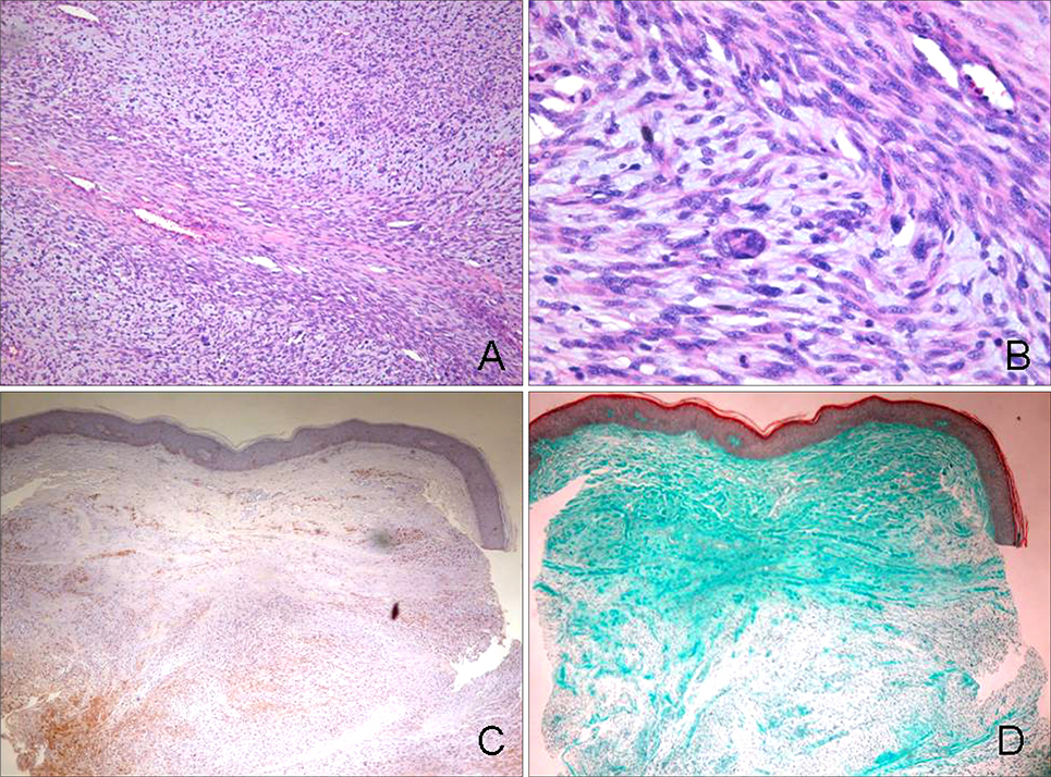
Fig. 2 The biopsy specimen showed a large mass in the dermis, showing a dense cellularity of spindle cells in a fascicular pattern (A: H&E, × 100). Upon magnification, compact bundles of spindle cells with pleomorphic, atypical nuclei were seen arranged in a whorling pattern. An anaplastic tumor giant cell is also seen (B: H&E, × 400). The tumor cells showed focal positivity to S-100 protein (C: S-100 protein, × 100), and a few weakly cyanophilic tumor cells were found upon Masson trichrome staining (D: Masson trichrome, × 100).

Fig. 3 PET-CT of the whole body showed an increase in FDG uptake in the left inguinal region (arrow), findings suggestive of lymph node metastasis, which was confirmed with biopsy.
Reference
-
1. McCormack LJ, Hazard JB, Dickson JA. Malignant epithelioid neurilemoma (schwannoma). Cancer. 1954; 7:725–728.
Article2. Ducatman BS, Scheithauer BW, Piepgras DG, Reiman HM, Ilstrup DM. Malignant peripheral nerve sheath tumors. A clinicopathologic study of 120 cases. Cancer. 1986; 57:2006–2021.
Article3. Choi DY, Park JW, Son SJ. A case of solitary malignant schwannoma with regional lymph node metastasis. Korean J Dermatol. 1981; 19:347–351.4. Chon HJ, Lee DK, Kim DJ, Son SJ. A case of purely epithelioid peripheral nerve sheath tumor. Korean J Dermatol. 2000; 38:518–521.5. Lee JH, Park CJ, Yi JY, Kim JS, Jeon HM. A case of malignant peripheral nerve sheath tumor in neurofibromatosis. Korean J Dermatol. 1999; 37:1535–1537.6. Lee JY, Suh JI, Ihm CW. Two cases of neurofibrosarcoma associated with multiple neurofibromatois. Korean J Dermatol. 1988; 26:110–115.7. Park HJ, Cinn YW. Malignant schwannoma. Korean J Dermatol. 1986; 24:159–163.8. Garg A, Gupta V, Gaikwad SB, Mishra NK, Ojha BK, Chugh M, et al. Scalp malignant peripheral nerve sheath tumor (MPNST) with bony involvement and new bone formation: case report. Clin Neurol Neurosurg. 2004; 106:340–344.
Article9. Al Akloby O, Bukhari IA, El-Shawarby M, Mulhim FA. Malignant peripheral nerve sheath tumor of the skin: case report. Am J Clin Dermatol. 2006; 7:201–203.10. Fukushima S, Kageshita T, Wakasugi S, Matsushita S, Kaguchi A, Ishihara T, et al. Giant malignant peripheral nerve sheath tumor of the scalp. J Dermatol. 2006; 33:865–868.
Article11. Baehring JM, Betensky RA, Batchelor TT. Malignant peripheral nerve sheath tumor: the clinical spectrum and outcome of treatment. Neurology. 2003; 61:696–698.12. Dabski C, Reiman HM, Muller SA. Neurofibrosarcoma of skin and subcutaneous tissues. Mayo Clin Proc. 1990; 65:164–172.
Article13. Hruban RH, Shiu MH, Senie RT, Woodruff JM. Malignant peripheral nerve sheath tumors of the buttock and lower extremity. A study of 43 cases. Cancer. 1990; 66:1253–1265.
Article14. Ha SJ, Park YM, Yi JY, Kim TY, Kom CW, Lee JY, et al. Malignant peripheral nerve sheath tumor arising from neurofibromatosis. Ann Dermatol. 1996; 8:153–157.
Article15. Kim HD, Shin HJ, Park YL, Whang KU, Baik SH, Park JS. A case of malignant peripheral nerve sheath tumor arising from neurofibromatosis during pregnancy. Korean J Dermatol. 2006; 44:75–78.16. Lee JH, Kim YC, Park HJ, Cinn YW. Two cases of neurofibrosarcoma arising from cutaneous neurofibroma in patients with neurofibromatosis. Korean J Dermatol. 1998; 36:924–927.
- Full Text Links
-
- Actions
-
Cited
- CITED
-
- Close
- Share
-
- Similar articles
-
- Malignant Peripheral Nerve Sheath Tumor Arising from Neurofibromatosis
- Malignant Peripheral Nerve Sheath Tumor of the Cauda Equina in Type I Neurofibromatosis: Case Report
- Malignant Peripheral Nerve Sheath Tumor Arising from Plexiform Neurofibroma in Neurofibromatosis Type 1
- A Case of Malignant Peripheral Nerve Sheath Tumor of the Neck Associated with Neurofibromatosis Type I
- Malignant Peripheral Nerve Sheath Tumor of the Larynx
