Rapid Growing Eosinophilic Granuloma in Skull after Minor Trauma
- Affiliations
-
- 1Department of Neurosurgery, The Catholic University of Korea, Bucheon St. Mary's Hospital, Bucheon, Korea. jkw94@naver.com
- KMID: 2156088
- DOI: http://doi.org/10.13004/kjnt.2015.11.1.22
Abstract
- The authors present a case of rapidly progressing eosinophilic granuloma (EG) of the skull without hemorrhage after minor trauma. A 6-year-old boy presented with a soft mass on the midline of his forehead. He had a surgery for EG 19 months ago. One month earlier, computed tomography (CT) and bone scans were performed to evaluate the possible recurrence of EG, and there was no evidence of recurrence in CT. However, a slightly increased uptake in the bone scan was noted on the midline of the forehead. A rapid growing mass developed in a new spot after a minor trauma 7 days before the patient arrived at the clinic. His physical examination was unremarkable, except for a non-tender, soft, and immobile mass. A plain skull X-ray and CT showed a lytic bony defect on the midline of the frontal bone. Magnetic resonance imaging showed a 1.4 cm sized enhancing mass. Surgical resection and cranioplasty were done. The role of trauma in the development of EG is unclear. However, our case suggests that minor trauma is an aggravating factor for EG formation. Careful observation with regular follow-up is necessary in patients with EG after minor trauma.
MeSH Terms
Figure
-
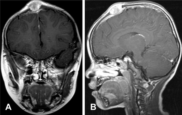
FIGURE 1 T1 enhanced images (A: coronal, B: sagittal) showing a 2.3 cm-sized enhancing mass in the mid vertex 19 months ago.
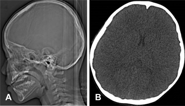
FIGURE 2 A: Skull lateral image taken one month prior to trauma showed no evidence of recurrence. B: Non-contrast axial computed tomography image.

FIGURE 3 A, B: Three-dimensional computed tomography showing lytic bony defect in the frontal region (small arrow: previous operation lesion, large arrow: new developed lesion). C, D: Sagital magnetic resonance imaging showing a 1.4 cm-sized enhnacing mass (C: non-contrast, D: contrast).
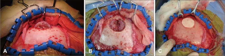
FIGURE 4 A: After the scalp flap was reflected, a protruding mass was found. (B) The mass was completely removed, and (C) a cranioplasty was performed with bone cement.
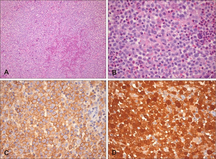
FIGURE 5 A, B: There are numerous oval Langerhans cells with many nuclei containing linear grooves (H-E, ×100). Eosinophilic microabscesses are noted (H-E, ×400). C: The surfaces of the Langerhans cells are uniformly positive for CD1a staining (×400). D: Strong positive staining is both nuclear and cytoplasmic with S-100 (×400).
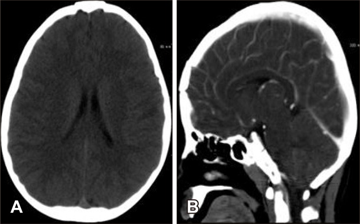
FIGURE 6 Computed tomography six months after the operation showing no recurrence. A: Axial noncontrast image. B: Sagittal contrast image.
Reference
-
1. Cho DY, Liau WR, Chiang IP. Eosinophilic granuloma with acute epidural hematoma: a case report. Pediatr Neurosurg. 2001; 35:266–269. PMID: 11741122.2. De Angulo G, Nair S, Lee V, Khatib Z, Ragheb J, Sandberg DI. Nonoperative management of solitary eosinophilic granulomas of the calvaria. J Neurosurg Pediatr. 2013; 12:1–5. PMID: 23662933.
Article3. Hasturk AE, Basmaci M. Multifocal extradural and intradural eosinophilic granuloma. J Craniofac Surg. 2013; 24:e214–e216. PMID: 23714965.
Article4. Jelsma F, Ross PJ. Tumor-like lesions of the calvaria (due to general disease processes). Am Surg. 1963; 29:247–255. PMID: 13964607.5. Kaul R, Gupta N, Gupta S, Gupta M. Eosinophilic granuloma of skull bone. J Cytol. 2009; 26:156–157. PMID: 21938183.
Article6. Lee KW, McLeary MS, Zuppan CW, Won DJ. Langerhans' cell histiocytosis presenting with an intracranial epidural hematoma. Pediatr Radiol. 2000; 30:326–328. PMID: 10836596.
Article7. Lee YS, Kwon JT, Park YS. Eosinophilic granuloma presenting as an epidural hematoma and cyst. J Korean Neurosurg Soc. 2008; 43:304–306. PMID: 19096637.
Article8. Manaka S, Izawa M, Nawata H. Skull tumor simulating sinus pericranii. Case report. J Neurosurg. 1977; 46:671–673. PMID: 845656.9. Martinez-Lage JF, Poza M, Cartagena J, Vicente JP, Biec F, de las Heras M. Solitary eosinophilic granuloma of the pediatric skull and spine. The role of surgery. Childs Nerv Syst. 1991; 7:448–451. PMID: 1790529.10. Mut M, Cataltepe O, Bakar B, Cila A, Akalan N. Eosinophilic granuloma of the skull associated with epidural haematoma: a case report and review of the literature. Childs Nerv Syst. 2004; 20:765–769. PMID: 15024599.
Article11. Sauerborn D, Pajić-Penavić I, Stojadinović T. Eosinophilic granuloma of the temporal bone in an adult: controversies in the management. Coll Antropol. 2012; 36(Suppl 2):163–166. PMID: 23397778.12. Vandenberg HJ, Coley BL. Primary tumors of the cranial bones. Surg Gynecol Obstet. 1950; 90:602–612. PMID: 15418430.
- Full Text Links
-
- Actions
-
Cited
- CITED
-
- Close
- Share
-
- Similar articles
-
- Eosinophilic Granuloma of the Proximal Humeral Epiphysis: A Case Report
- Eosinophilic Granuloma Presenting as an Epidural Hematoma and Cyst
- Aneurysmal Bone Cyst(ABC) Secondary to Eosinophilic Granuloma of the Skull: Case Report
- Eosinophilic Granuloma of Lumbar Spine in Old Patient: 1 Case Report
- Monostotic Eosinophilic Granuloma of the Skull: A Case Report
