Pyogenic Intradural Abscess of Lumbar Spine: A Case Report
- Affiliations
-
- 1Division of Pediatric Radiology, Seoul National University Children's Hospital, Seoul, Korea.
- 2Department of Neurosurgery, Seoul National University Boramae Hospital, Seoul, Korea. nsyang@brm.co.kr
- 3Department of Neurosurgery, Jeju National University Children's Hospital, Jeju, Korea.
- KMID: 2156087
- DOI: http://doi.org/10.13004/kjnt.2015.11.1.18
Abstract
- We report a case of spinal intradural abscess which shows serial changes on magnetic resonance imaging (MRI). Well-encapsulated, rim-enhancing lesion with mass effect was visualized at ventral side of lumbar spinal canal on 17 days after initial negative MRI, which was thought to be epidural abscess. It was revealed to be intradural in location on operation and successfully treated by drainage and antibiotics. Follow-up MRI showed resolution of abscess. Clinical significance and pathogenesis of this case was briefly discussed.
MeSH Terms
Figure
-
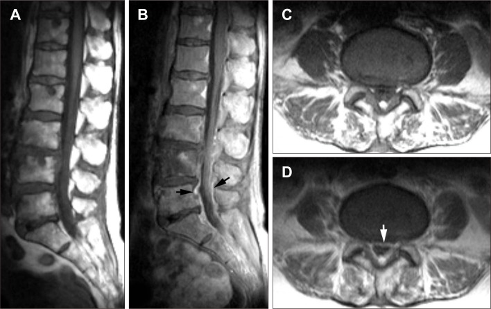
FIGURE 1 Initial magnetic resonance imaging of the lumbar spine. Precontrast T1-weighted images (A, C) and postcontrast T1-weighted images (B, D) depict irregular, linear enhancement in the epidural venous plexus at L4-5 and L5-S1 level (black arrows in B, white arrow in D). There was no abscess formation.
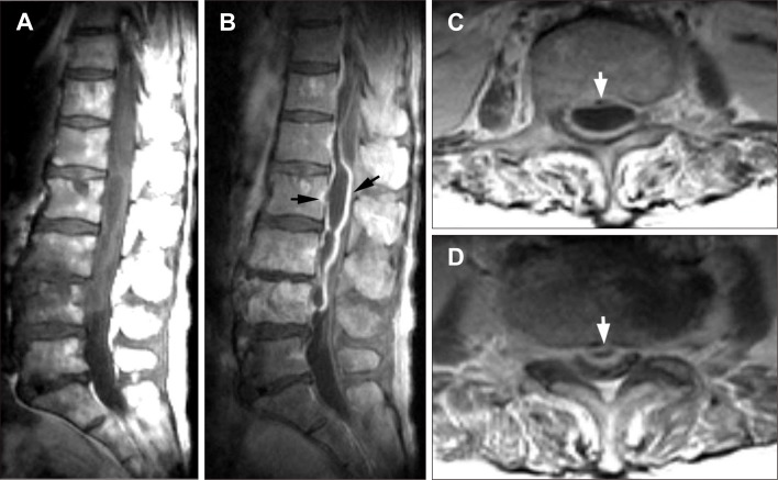
FIGURE 2 Preoperative follow-up magnetic resonance imaging obtained on 17th day of intravenous antibiotic treatment. Precontrast T1-weighted images (A) and postcontrast T1-weighted images (B-D) show elongated shape fluid collection with rim-enhancement at ventral side of spinal canal (arrows) at L2-4 level. On axial images at L2 (C) and L3-4 level (D) the abscess shows dorsally convex and mass effect on the dural sac mimicking epidural lesion (arrows).

FIGURE 3 Intraoperative photographs. A: Yellowish purulent material was oozing out (black arrow) between lumbar nerve roots (white asterisks) after opening of cyst wall. B: A part of cyst wall was grasped by forceps.
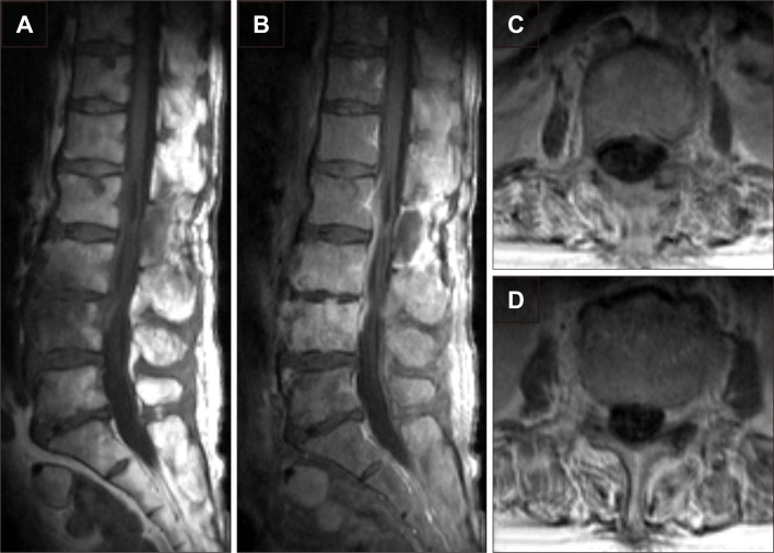
FIGURE 4 Follow-up magnetic resonance imaging (MRI) obtained 2 weeks after operation. Precontrast T1-weighted images (A) and postcontrast T1-weighted images (B-D) show near complete disappearance of intraspinal rim-enhancing lesion observed on preoperative MRI as described in Figure 2.
Reference
-
1. Agarwal N, Shah J, Hansberry DR, Mammis A, Sharer LR, Goldstein IM. Presentation of cauda equina syndrome due to an intradural extramedullary abscess: a case report. Spine J. 2014; 14:e1–e6. PMID: 24331844.
Article2. Bartels RH, de Jong TR, Grotenhuis JA. Spinal subdural abscess. Case report. J Neurosurg. 1992; 76:307–311. PMID: 1346157.3. Brecker SJ, Pugey CD. Nocardia asteroides infection of the cauda equina. J Neurol Neurosurg Psychiatry. 1988; 51:309–311. PMID: 3346704.
Article4. Dacey RG, Winn HR, Jane JA, Butler AB. Spinal subdural empyema: report of two cases. Neurosurgery. 1978; 3:400–403. PMID: 154072.5. Fraser RA, Ratzan K, Wolpert SM, Weinstein L. Spinal subdural empyema. Arch Neurol. 1973; 28:235–238. PMID: 4688429.
Article6. Hadjipavlou AG, Mader JT, Necessary JT, Muffoletto AJ. Hematogenous pyogenic spinal infections and their surgical management. Spine (Phila Pa 1976). 2000; 25:1668–1679. PMID: 10870142.
Article7. Hasan MY, Kumar KK, Lwin S, Lau LL, Kumar N. Cervical intradural abscess masquerading as an epidural collection. Global Spine J. 2013; 3:249–252. PMID: 24436877.
Article9. Inoue H, Hirai T, Nagaya T, Takeda F, Kawafuchi J. [Spinal subdural abscess-report of a case (author's transl)]. No Shinkei Geka. 1977; 5:169–172. PMID: 557740.10. Kurokawa Y, Hashi K, Fujishige M, Tsuchida M, Maeda K, Kaneko M. [Spinal subdural empyema diagnosed by MRI and recovered by conservative treatment]. No To Shinkei. 1989; 41:513–517. PMID: 2572246.11. Levy ML, Wieder BH, Schneider J, Zee CS, Weiss MH. Subdural empyema of the cervical spine: clinicopathological correlates and magnetic resonance imaging. Report of three cases. J Neurosurg. 1993; 79:929–935. PMID: 7902429.12. Lim HY, Choi HJ, Kim S, Kuh SU. Chronic spinal subdural abscess mimicking an intradural-extramedullary tumor. Eur Spine J. 2013; 22(Suppl 3):S497–S500. PMID: 23397217.
Article13. Martin RJ, Yuan HA. Neurosurgical care of spinal epidural, subdural, and intramedullary abscesses and arachnoiditis. Orthop Clin North Am. 1996; 27:125–136. PMID: 8539043.
Article14. Sadato N, Numaguchi Y, Rigamonti D, Kodama T, Nussbaum E, Sato S, et al. Spinal epidural abscess with gadolinium-enhanced MRI: serial follow-up studies and clinical correlations. Neuroradiology. 1994; 36:44–48. PMID: 8107997.
Article15. Sathi S, Schwartz M, Cortez S, Rossitch E Jr. Spinal subdural abscess: successful treatment with limited drainage and antibiotics in a patient with AIDS. Surg Neurol. 1994; 42:424–427. PMID: 7974149.
Article16. Thomé C, Krauss JK, Zevgaridis D, Schmiedek P. Pyogenic abscess of the filum terminale. Case report. J Neurosurg. 2001; 95(1 Suppl):100–104. PMID: 11453406.
- Full Text Links
-
- Actions
-
Cited
- CITED
-
- Close
- Share
-
- Similar articles
-
- Pediatric Lumbar Epidural Abscess Combined with Cauda Equina Syndrome: Case Report
- Pyogenic Arthritis of the Facet Joint with Concurrent Epidural and Paraspinal Abscess: A Case Report
- Pyogenic Spinal Epidural Abscess: A Case Report
- Huge Intradural Lumbar Disc Herniation Mimicking an Intradural Spinal Tumor: A Case Report
- Pyogenic L4-5 Spondylitis Managed with Percutaneous Drainage Followed by Posterior Lumbar Interbody Fusion: A Case Report
