Spontaneous Extracranial Vertebral Artery Dissection in a Neurofibromatosis 1 Patient with Bilateral Intrathoracic Spinal Meningoceles around the Scoliosis: Report of an Autopsy Case
- Affiliations
-
- 1Department of Forensic Investigation, National Forensic Service Seoul Institute, Seoul, Korea. jhjh7176@korea.kr
- KMID: 2155725
- DOI: http://doi.org/10.7580/kjlm.2016.40.1.14
Abstract
- Neurofibromatosis 1 (NF1) is a common autosomal dominant disorder that causes several systemic diseases. Many studies have reported that NF1 is associated with intrathoracic meningoceles and scoliosis. The incidence of vertebral artery dissection is estimated to be 1-1.5 per 100,000 population. We experienced an autopsy case of massive intrathoracic hemorrhage due to spontaneous vertebral artery dissection in a patient with NF1, who had intrathoracic spinal meningoceles and scoliosis. A 47-year-old man was found dead at his home in the morning. He had a history of NF1 including numerous cutaneous neurofibromas and hyperpigmented macules, scoliosis, and deformity of the leg. The autopsy revealed the dissection and rupture of the left vertebral artery, and a pseudocyst that had formed due to arterial leakage on the wall of the meningocele on the left side. The pseudocyst had eventually ruptured and leaked blood, resulting in a massive hemothorax on the left side. Thus, it was revealed that the patient had suffered from NF1-associated intrathoracic meningoceles and scoliosis, and we concluded that the cause of his death was a massive hemothorax on the left side, caused by the dissection and rupture of the left vertebral artery.
MeSH Terms
Figure
-
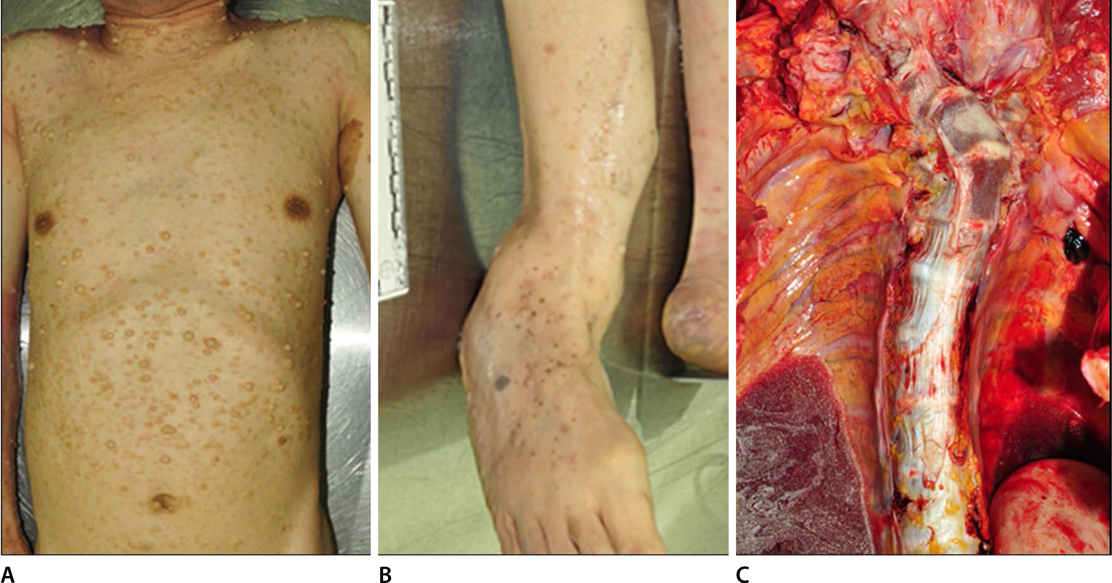
Fig. 1. External and internal autopsy findings consistent with neurofibromatosis. (A) Numerous cutaneous neurofibromas and hyperpigmented macules were noticed in the patient's body. (B) Skeletal deformity of the right leg and scars from a previous operation were observed. (C) Scoliosis was observed at the cervical and thoracic spine.
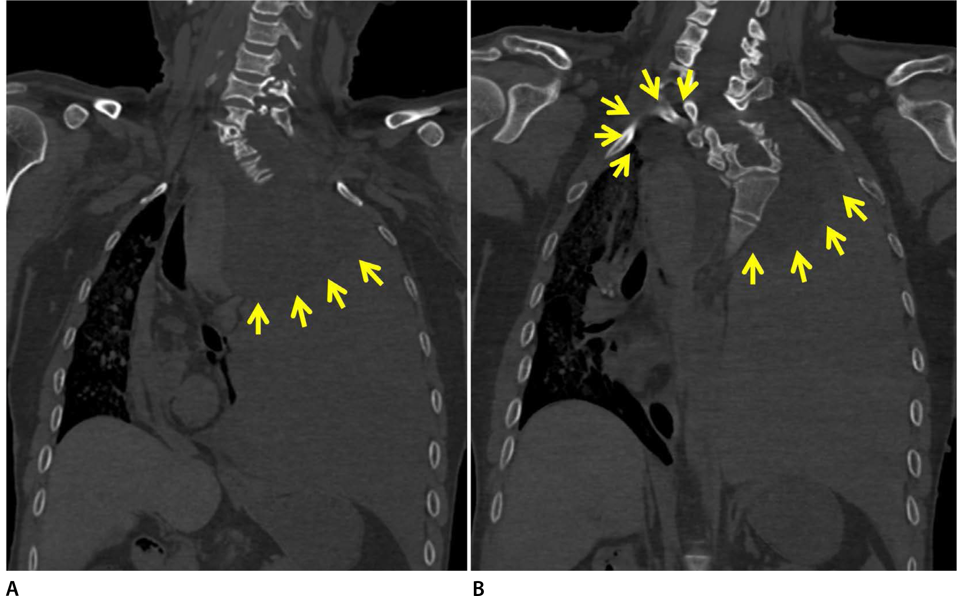
Fig. 2. Postmortem computed tomography findings. (A) A ‘white out’ of the left thorax with a mediastinal shift to the right was revealed. (A, B) Bilateral round masses were seen at the superior intrathoracic area (masses are indicated by yellow arrows).

Fig. 3. Intrathoracic meningoceles found upon internal examination. (A) Blood had leaked out of the round mass in the left intrathoracic area. (B, C) The left intrathoracic round mass was revealed to be a spinal meningocele filled with clear cerebrospinal fluid. A pseudocyst filled with blood and hematoma was found at the wall of the intrathoracic meningocele on left side. (D) The bilateral intrathoracic round masses at the bilateral T2-T3 intervertebral foramen were revealed to be spinal meningoceles (bilateral meningoceles are indicated by yellow arrows).
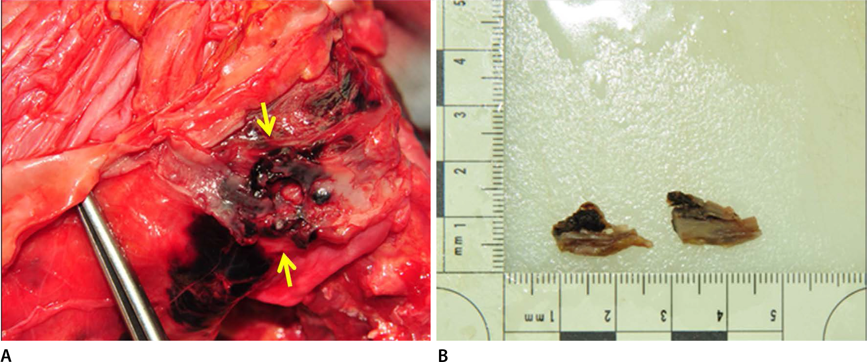
Fig. 4. (A, B) Upon close examination, dissection and rupture of the prevertebral segment of the left vertebral artery were noticed (the ruptured areas are indicated by yellow arrows).
Reference
-
1.Lee VH., Brown RD Jr., Mandrekar JN, et al. Incidence and outcome of cervical artery dissection: a population-based study. Neurology. 2006. 67:1809–12.
Article2.Haneline MT., Rosner AL. The etiology of cervical artery dissection. J Chiropr Med. 2007. 6:110–20.
Article3.Kumar V., Abbas AK., Aster JC. Robbins and Cotran pathologic basis of disease. 9th ed.Philadelphia, PA: Elsevier/Saunders;2015.4.Ueda K., Honda O., Satoh Y, et al. Computed tomography (CT) findings in 88 neurofibromatosis 1 (NF1) patients: Prevalence rates and correlations of thoracic findings. Eur J Radiol. 2015. 84:1191–5.
Article5.Swetz KM., Spinner RJ. Large intrathoracic meningocele associated with neurofibromatosis type 1. Mayo Clin Proc. 2009. 84:769.
Article6.Na JY., Park JP., Kim DW, et al. Aneurysmal rupture of the internal carotid artery in a presumed neurofibromatosis type I patient. Korean J Leg Med. 2013. 37:34–7.
Article7.Miura H., Taira O., Uchida O, et al. Spontaneous haemothorax associated with von Recklinghausen's disease: review of occurrence in Japan. Thorax. 1997. 52:577–8.
Article8.Fohrding LZ., Sellmann T., Angenendt S, et al. A case of lethal spontaneous massive hemothorax in a patient with neurofibromatosis 1. J Cardiothorac Surg. 2014. 9:172.
Article9.Uranishi R., Ochiai C., Okuno S, et al. Cerebral aneurysms associated with von Recklinghausen neurofibromatosis: report of two cases. No Shinkei Geka. 1995. 23:237–42.10.Sobata E., Ohkuma H., Suzuki S. Cerebrovascular disorders associated with von Recklinghausen's neurofibromatosis: a case report. Neurosurgery. 1988. 22:544–9.
Article
- Full Text Links
-
- Actions
-
Cited
- CITED
-
- Close
- Share
-
- Similar articles
-
- A Case of Bilateral Spontaneous Extracranial Vertebral Artery Dissection
- Giant Intrathoracic Meningocele and Breast Cancer in a Neurofibromatosis Type I Patient
- Endovascular Treatment for Spontaneous Vertebral Arteriovenous Fistula in Neurofibromatosis Type 1: A Case Report
- Extracranial vertebral arteriovenous fistula presenting as an osteolytic lesion of the axis: Case report
- Intracranial and Extracranial Fusiform Aneurysms in a Patient with Neurofibromatosis Type 1: A Case Report