Korean J Radiol.
2015 Aug;16(4):874-880. 10.3348/kjr.2015.16.4.874.
Diffusion-Weighted Imaging for Pretreatment Evaluation and Prediction of Treatment Effect in Patients Undergoing CT-Guided Injection for Lumbar Disc Herniation
- Affiliations
-
- 1Department of Radiology, Affiliated Hospital of Chengdu University, Chengdu, Sichuan Province 610000, China.
- 2Sichuan Key Laboratory of Medical Imaging and Department of Radiology, Affiliated Hospital of North Sichuan Medical College, Nanchong, Sichuan Province 637000, China. niu19850519@163.com
- KMID: 2155562
- DOI: http://doi.org/10.3348/kjr.2015.16.4.874
Abstract
OBJECTIVE
To determine whether a change in apparent diffusion coefficient (ADC) value could predict early response to CT-guided Oxygen-Ozone (O2-O3) injection therapy in patients with unilateral mono-radiculopathy due to lumbar disc herniation.
MATERIALS AND METHODS
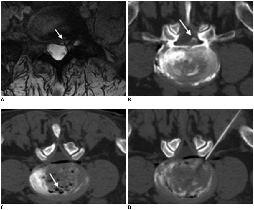
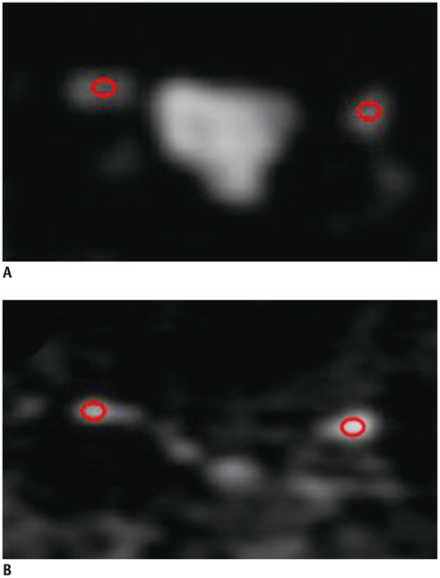
A total of 52 patients with unilateral mono-radiculopathy received a single intradiscal (3 mL) and periganglionic (5 mL) injection of an O2-O3 mixture. An ADC index of the involved side to the intact side was calculated using the following formula: pre-treatment ADC index = ([ADC involved side - ADC intact side] / ADC intact side) x 100. We analyzed the relationship between the pre-treatment Oswestry Disability Index (ODI) and the ADC index. In addition, the correlation between ODI recovery ratio and ADC index was investigated. The sensitivity and specificity of the ADC index for predicting response in O2-O3 therapy was determined.
RESULTS
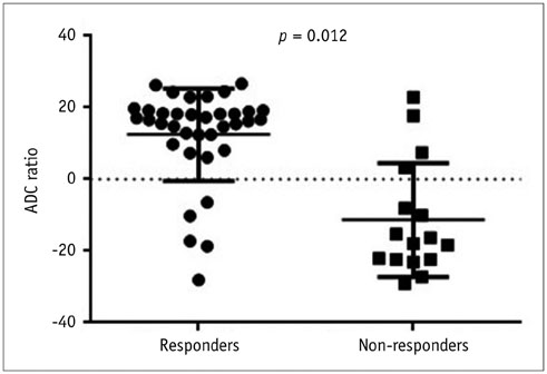
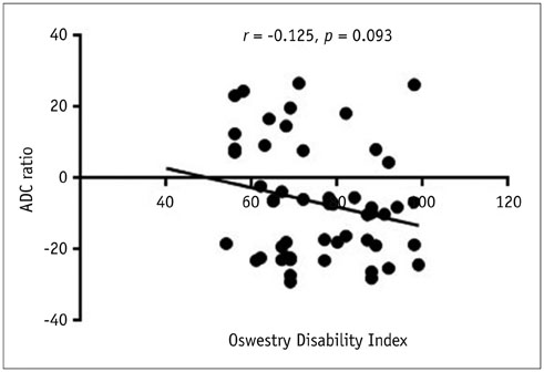
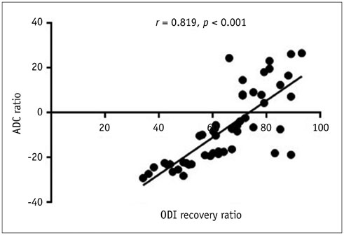
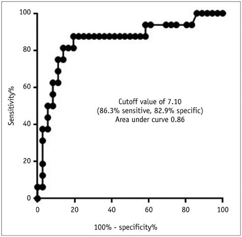
Oswestry Disability Index and the ADC index was not significantly correlated (r = -0.125, p = 0.093). The ADC index and ODI recovery ratio was significantly correlated (r = 0.819, p < 0.001). When using 7.10 as the cut-off value, the ADC index obtained a sensitivity of 86.3% and a specificity of 82.9% for predicting successful response to therapy around the first month of follow-up.
CONCLUSION
This preliminary study demonstrates that the patients with decreased ADC index tend to show poor improvement of clinical symptoms. The ADC index may be a useful indicator to predict early response to CT-guided O2-O3 injection therapy in patients with unilateral mono-radiculopathy due to lumbar disc herniation.
Keyword
MeSH Terms
-
Adult
Aged
Diffusion Magnetic Resonance Imaging/*methods
Female
Humans
Intervertebral Disc Displacement/*diagnosis/*therapy
Lumbar Vertebrae/*pathology
Male
Middle Aged
Oxygen/therapeutic use
Ozone/therapeutic use
Sensitivity and Specificity
Tomography, X-Ray Computed/*methods
Treatment Outcome
Young Adult
Oxygen
Ozone
Figure
Reference
-
1. Andreula CF, Simonetti L, De Santis F, Agati R, Ricci R, Leonardi M. Minimally invasive oxygen-ozone therapy for lumbar disk herniation. AJNR Am J Neuroradiol. 2003; 24:996–1000.2. Bocci V. Biological and clinical effects of ozone. Has ozone therapy a future in medicine? Br J Biomed Sci. 1999; 56:270–227.3. Lehnert T, Mundackatharappel S, Schwarz W, Bisdas S, Wetter A, Herzog C, et al. [Nucleolysis in the herniated disk]. Radiologe. 2006; 46:513–519.4. Oder B, Loewe M, Reisegger M, Lang W, Ilias W, Thurnher SA. CT-guided ozone/steroid therapy for the treatment of degenerative spinal disease--effect of age, gender, disc pathology and multi-segmental changes. Neuroradiology. 2008; 50:777–785.5. Lehnert T, Naguib NN, Wutzler S, Nour-Eldin NE, Bauer RW, Kerl JM, et al. Analysis of disk volume before and after CT-guided intradiscal and periganglionic ozone-oxygen injection for the treatment of lumbar disk herniation. J Vasc Interv Radiol. 2012; 23:1430–1436.6. Komori H, Shinomiya K, Nakai O, Yamaura I, Takeda S, Furuya K. The natural history of herniated nucleus pulposus with radiculopathy. Spine (Phila Pa 1976). 1996; 21:225–222.7. Splendiani A, Puglielli E, De Amicis R, Barile A, Masciocchi C, Gallucci M. Spontaneous resolution of lumbar disk herniation: predictive signs for prognostic evaluation. Neuroradiology. 2004; 46:916–922.8. Splendiani A, Perri M, Conchiglia A, Fasano F, Di Egidio G, Masciocchi C, et al. MR assessment of lumbar disk herniation treated with oxygen-ozone diskolysis: the role of DWI and related ADC versus intervertebral disk volumetric analysis for detecting treatment response. Neuroradiol J. 2013; 26:347–356.9. Basser PJ, Jones DK. Diffusion-tensor MRI: theory, experimental design and data analysis - a technical review. NMR Biomed. 2002; 15:456–467.10. Beaulieu C, Does MD, Snyder RE, Allen PS. Changes in water diffusion due to Wallerian degeneration in peripheral nerve. Magn Reson Med. 1996; 36:627–631.11. Basser PJ, Pierpaoli C. Microstructural and physiological features of tissues elucidated by quantitative-diffusion-tensor MRI. 1996. J Magn Reson. 2011; 213:560–557.12. Hiltunen J, Suortti T, Arvela S, Seppä M, Joensuu R, Hari R. Diffusion tensor imaging and tractography of distal peripheral nerves at 3 T. Clin Neurophysiol. 2005; 116:2315–2323.13. Khalil C, Hancart C, Le Thuc V, Chantelot C, Chechin D, Cotten A. Diffusion tensor imaging and tractography of the median nerve in carpal tunnel syndrome: preliminary results. Eur Radiol. 2008; 18:2283–2291.14. Kabakci N, Gürses B, Firat Z, Bayram A, Uluğ AM, Kovanlikaya A, et al. Diffusion tensor imaging and tractography of median nerve: normative diffusion values. AJR Am J Roentgenol. 2007; 189:923–927.15. Chen YY, Lin XF, Zhang F, Zhang X, Hu HJ, Wang DY, et al. Diffusion tensor imaging of symptomatic nerve roots in patients with cervical disc herniation. Acad Radiol. 2014; 21:338–344.16. Fairbank JC, Couper J, Davies JB, O'Brien JP. The Oswestry low back pain disability questionnaire. Physiotherapy. 1980; 66:271–273.17. Gallucci M, Limbucci N, Zugaro L, Barile A, Stavroulis E, Ricci A, et al. Sciatica: treatment with intradiscal and intraforaminal injections of steroid and oxygen-ozone versus steroid only. Radiology. 2007; 242:907–913.18. Borich MR, Wadden KP, Boyd LA. Establishing the reproducibility of two approaches to quantify white matter tract integrity in stroke. Neuroimage. 2012; 59:2393–2400.19. Kunogi J, Hasue M. Diagnosis and operative treatment of intraforaminal and extraforaminal nerve root compression. Spine (Phila Pa 1976). 1991; 16:1312–1132.20. Hasegawa T, Mikawa Y, Watanabe R, An HS. Morphometric analysis of the lumbosacral nerve roots and dorsal root ganglia by magnetic resonance imaging. Spine (Phila Pa 1976). 1996; 21:1005–1100.21. Jenis LG, An HS. Spine update. Lumbar foraminal stenosis. Spine (Phila Pa 1976). 2000; 25:389–339.22. Muto M, Andreula C, Leonardi M. Treatment of herniated lumbar disc by intradiscal and intraforaminal oxygen-ozone (O2-O3) injection. J Neuroradiol. 2004; 31:183–189.23. Muto M, Avella F. Percutaneous treatment of herniated lumbar disc by intradiscal oxygen-ozone injection. Interv Neuroradiol. 1998; 4:279–286.24. Zhang Y, Ma Y, Jiang J, Ding T, Wang J. Treatment of the lumbar disc herniation with intradiscal and intraforaminal injection of oxygen-ozone. J Back Musculoskelet Rehabil. 2013; 26:317–322.25. Lutze M, Stendel R, Vesper J, Brock M. Periradicular therapy in lumbar radicular syndromes: methodology and results. Acta Neurochir (Wien). 1997; 139:719–772.26. Takashima H, Takebayashi T, Yoshimoto M, Terashima Y, Ida K, Yamashita T. Efficacy of diffusion-weighted magnetic resonance imaging in diagnosing spinal root disorders in lumbar disc herniation. Spine (Phila Pa 1976). 2013; 38:E998–E100.27. Mac Donald CL, Dikranian K, Bayly P, Holtzman D, Brody D. Diffusion tensor imaging reliably detects experimental traumatic axonal injury and indicates approximate time of injury. J Neurosci. 2007; 27:11869–11876.28. Morisaki S, Kawai Y, Umeda M, Nishi M, Oda R, Fujiwara H, et al. In vivo assessment of peripheral nerve regeneration by diffusion tensor imaging. J Magn Reson Imaging. 2011; 33:535–542.29. Ford JC, Hackney DB, Alsop DC, Jara H, Joseph PM, Hand CM, et al. MRI characterization of diffusion coefficients in a rat spinal cord injury model. Magn Reson Med. 1994; 31:488–494.30. Sakai K, Yamada K, Oouchi H, Nishimura T. Numerical simulation model of hyperacute/acute stage white matter infarction. Magn Reson Med Sci. 2008; 7:187–194.31. Chang KJ, Kamel IR, Macura KJ, Bluemke DA. 3.0-T MR imaging of the abdomen: comparison with 1.5 T. Radiographics. 2008; 28:1983–1998.32. Olsrud J, Lätt J, Brockstedt S, Romner B, Björkman-Burtscher IM. Magnetic resonance imaging artifacts caused by aneurysm clips and shunt valves: dependence on field strength (1.5 and 3 T) and imaging parameters. J Magn Reson Imaging. 2005; 22:433–443.33. Wiesinger F, Van de Moortele PF, Adriany G, De Zanche N, Ugurbil K, Pruessmann KP. Potential and feasibility of parallel MRI at high field. NMR Biomed. 2006; 19:368–378.34. Miyoshi S, Sekiguchi M, Konno S, Kikuchi S, Kanaya F. Increased expression of vascular endothelial growth factor protein in dorsal root ganglion exposed to nucleus pulposus on the nerve root in rats. Spine (Phila Pa 1976). 2011; 36:E1–EE.35. Kobayashi S, Uchida K, Kokubo Y, Takeno K, Yayama T, Miyazaki T, et al. Synapse involvement of the dorsal horn in experimental lumbar nerve root compression: a light and electron microscopic study. Spine (Phila Pa 1976). 2008; 33:716–772.
- Full Text Links
- Actions
-
Cited
- CITED
-
- Close
- Share
- Similar articles
-
- The Relationship between the Lower Lumbar Disc Herniation and the Morphology of the Iliolumbar Ligaments Using Magnetic Resonance Imaging
- Classification and Imaging Study of the Lumbar Disc Herniation
- Lumbar Epidural Venography in the Diagnosis of Lumbar Disc Herniation
- Clinical Analysis of Recurrent Lumbar Disc Herniation
- The Role of CT Discography in Far Lateral Disk Herniation

