Korean J Radiol.
2015 Jun;16(3):662-667. 10.3348/kjr.2015.16.3.662.
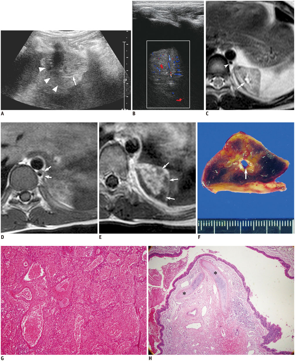
Extralobar Pulmonary Sequestration with Hemorrhagic Infarction in a Child: Preoperative Imaging Diagnosis and Pathological Correlation
- Affiliations
-
- 1Department of Radiology and Research Institute of Radiology, Asan Medical Center, University of Ulsan College of Medicine, Seoul 138-736, Korea. hwgoo@amc.seoul.kr
- KMID: 2155538
- DOI: http://doi.org/10.3348/kjr.2015.16.3.662
Abstract
- We describe a rare case of extralobar pulmonary sequestration with hemorrhagic infarction in a 10-year-old boy who presented with acute abdominal pain and fever. In our case, internal branching linear architecture, lack of enhancement in the peripheral portion of the lesion with internal hemorrhage, and vascular pedicle were well visualized on preoperative magnetic resonance imaging that led to successful preoperative diagnosis of extralobar pulmonary sequestration with hemorrhagic infarction probably due to torsion.
Keyword
MeSH Terms
Figure
Reference
-
1. Chen W, Wagner L, Boyd T, Nagarajan R, Dasgupta R. Extralobar pulmonary sequestration presenting with torsion: a case report and review of literature. J Pediatr Surg. 2011; 46:2025–2028.2. Gawlitza M, Hirsch W, Weißer M, Ritter L, Metzger RP. Torsion of extralobar lung sequestration - lack of contrast medium enhancement could facilitate MRI-based diagnosis. Klin Padiatr. 2014; 226:38–39.3. Huang EY, Monforte HL, Shaul DB. Extralobar pulmonary sequestration presenting with torsion. Pediatr Surg Int. 2004; 20:218–220.4. Kirkendall ES, Guiot AB. Torsed pulmonary sequestration presenting with gastrointestinal manifestations. Clin Pediatr (Phila). 2013; 52:981–984.5. Lima M, Randi B, Gargano T, Tani G, Pession A, Gregori G. Extralobar pulmonary sequestration presenting with torsion and associated hydrothorax. Eur J Pediatr Surg. 2010; 20:208–210.6. Mammen A, Myers NA, Beasley SW. Torsion and infarction of an extralobar pulmonary sequestration. Pediatr Surg Int. 1994; 9:399–400.7. Shah R, Carver TW, Rivard DC. Torsed pulmonary sequestration presenting as a painful chest mass. Pediatr Radiol. 2010; 40:1434–1435.8. Uchida DA, Moore KR, Wood KE, Pysher TJ, Downey EC. Infarction of an extralobar pulmonary sequestration in a young child: diagnosis and excision by video-assisted thoracoscopy. J Laparoendosc Adv Surg Tech A. 2010; 20:399–401.9. Zucker EJ, Tracy DA, Chwals WJ, Solky AC, Lee EY. Paediatric torsed extralobar sequestration containing calcification: Imaging findings with pathological correlation. Clin Radiol. 2013; 68:94–97.10. Takeuchi K, Ono A, Yamada A, Toyooka M, Takahashi T, Shigematsu Y, et al. Two adult cases of extralobar pulmonary sequestration: a non-complicated case and a necrotic case with torsion. Pol J Radiol. 2014; 79:145–149.
- Full Text Links
- Actions
-
Cited
- CITED
-
- Close
- Share
- Similar articles
-
- Congenital Cystic Adenomatoid Malformation Associated with Extralobar Pulmonary Sequestration: A case report
- Unusual Presentation of Extralobar Pulmonary Sequestration: A Case Report
- Extralobar Pulmonary Sequestration located in Right Oblique Fissure with Unusual Vascularture
- Intraabdominal Extralobar Pulmonary Sequestration Detected by Prenatal Ultrasound
- A case of pulmonary sequestration mimicking mediastinal mass detected by prenatal ultrasonography


