Knotted stents: Case report and outcome analysis
- Affiliations
-
- 1Department of Urology, Seoul Medical Center, Seoul, Korea. cau1004@naver.com
- 2Department of Urology, Senam Hospital, Seoul, Korea.
- 3Department of Radiology, Seoul Medical Center, Seoul, Korea.
- KMID: 2155322
- DOI: http://doi.org/10.4111/kju.2015.56.5.405
Abstract
- A knotted ureteral stent is an extremely rare condition, with fewer than 20 cases reported in the literature; however, it is difficult to treat. We report a case in which a folded Terumo guidewire was successfully used to remove a knotted stent percutaneously without anesthesia. We also review the current literature on predisposing factors and management strategies for knotted ureteral stents.
Keyword
MeSH Terms
Figure
-
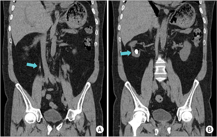
Fig. 1 Coronal scans obtained via nonenhanced computed tomography. A 0.4-cm upperureteral stone with no hydronephrosis (A: blue arrow) and a 2.0-cm renal stone in lower calyx (B: blue arrow) were identified.
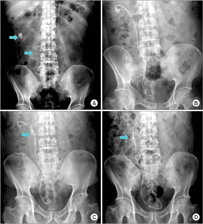
Fig. 2 X-ray images of the kidney, ureter, and bladder. (A) Pretreatment (upper arrow: renal stone, lower arrow: upper ureteral stone). (B) Immediately after insertion of a double-J stent. (C) Steinstrasse after extracorporeal shock wave lithotripsy (blue arrow: steinstrasse). (D) Knot formation (blue arrow: knott).
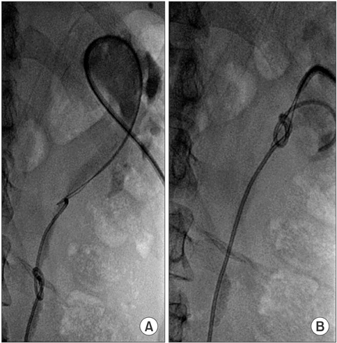
Fig. 3 Fluoroscopic images of antegrade ureteral stent removal. A 7-French angio sheath was inserted under ultrasonographic and fluoroscopic guidance, and the KMP (Beacon Tip Torcon NB advantage angiographic catheter, Cook Medical, Bloomington, IN, USA) catheter was inserted through the sheath. (A) A folded 0.018 Terumo guidewire was inserted in the area of the knot formation through the KMP catheter. (B) The knotted stent was caught in the folded guidewire and was removed percutaneously. After the procedure, an 8.5-French pigtail catheter was inserted in the kidney.
Reference
-
1. Zimskind PD, Fetter TR, Wilkerson JL. Clinical use of long-term indwelling silicone rubber ureteral splints inserted cystoscopically. J Urol. 1967; 97:840–844.2. Ahallal Y, Khallouk A, El Fassi MJ, Farih MH. Risk factor analysis and management of ureteral double-j stent complications. Rev Urol. 2010; 12:e147–e151.3. Groeneveld AE. The role of ESWL in the treatment of large kidney stones. Singapore Med J. 1989; 30:249–254.4. Picozzi S, Carmignani L. A knotted ureteral stent: A case report and review of the literature. Urol Ann. 2010; 2:80–82.5. Rivalta M, Sighinolfi MC, Micali S, De Stefani S, Bianchi G. Knotted ureteral catheter in an 83-year-old man: case presentation and urological non-invasive management in the elderly. Urol Res. 2009; 37:261–262.6. Moufid K, Touiti D, Mohamed L. "Knot stent": an unusual cause of acute renal failure in solitary kidney. J Clin Imaging Sci. 2012; 2:36.7. Nettle J, Huang JG, Rao R, Costello AJ. Ureteroscopic holmium laser ablation of a knotted ureteral stent. J Endourol. 2012; 26:968–970.8. Bhirud P, Giridhar V, Hegde P. Midureteric knotted stent removed by percutaneous access. Urol Ann. 2012; 4:106–107.
- Full Text Links
-
- Actions
-
Cited
- CITED
-
- Close
- Share
-
- Similar articles
-
- Removal of Knotted Nasogastric Tube: A Literature Review and Lessons From Our Case
- Intracardiac Knotting of a Balloon-tipped, Flow-directed Pulmonary Artery Catheter: A Case Report
- Upward Migration of Distal Ventriculoperitoneal Shunt Catheter into the Heart : Case Report
- Surgical Removal of Knotted Pulmonary Artery Catheter: A Case Report
- A double-knotted pulmonary artery catheter with large loop in the right internal jugular vein: A case report
