Right sided double inferior vena cava with obstructed retrocaval ureter: Managed with single incision multiple port laparoscopic technique using "Santosh Postgraduate Institute tacking ureteric fixation technique"
- Affiliations
-
- 1Department of Urology, Postgraduate Institute of Medical Education and Research (PGIMER), Chandigarh, India. santoshsp1967jaimatadi@yahoo.co.in
- KMID: 2155310
- DOI: http://doi.org/10.4111/kju.2015.56.4.330
Abstract
- Right double inferior vena cava with obstructed retrocaval ureter is an extremely rare anomaly with only a few reported cases in the literature. To the best of our knowledge, this is the first case report describing ureteric repair by use of a single-incision laparoscopic technique. In addition, this report addresses the underlying surgical challenges of this repair and provides a brief review of the embryology of this anomaly. The "Santosh Postgraduate Institute ureteric tacking fixation technique" provides ease of end-to-end uretero-ureteric anastomosis in a single-incision laparoscopic surgery.
Keyword
MeSH Terms
-
Humans
Intraoperative Care/methods
Intraoperative Complications/*prevention & control
Laparoscopy/methods
Magnetic Resonance Imaging
Male
*Retrocaval Ureter/diagnosis/physiopathology/surgery
Treatment Outcome
Urography/methods
Urologic Surgical Procedures/*methods
*Vena Cava, Inferior/abnormalities/surgery
Young Adult
Figure
-

Fig. 1 (A) Intraoperative retrograde pyelography showing the characteristic fish-hook sign of right retrocaval ureter. (B) Magnetic resonance imaging (MRI): preoperative image showing right retrocaval ureter with right double inferior vena cava (IVC). (C) Postoperative MRI showing normal caliber right ureter following repair with SIMPLE technique. SIMPLE, single incision multiple port laparo-endoscpic.
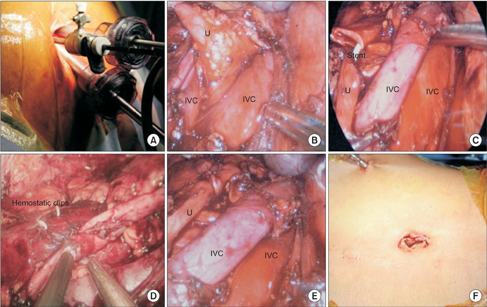
Fig. 2 (A) Port placement in SIMPLE technique. (B) Intraoperative image showing ureter with periureteric tissue being mobilized between the right double inferior vena cava. (C) Both the ends of ureter brought to one side with excision of stenotic segment. (D) End-to-end uretero-ureteric anastomosis using "Santosh Postgraduate Institute tacking ureteric fixation". (E) Completed anastomosis. (F) Skin incision after closure. IVC, inferior vena cava; U, ureter.
Reference
-
1. Kumar S, Shankaregowda SA, Devana SK, Jain S, Singh SK. Single-incision multiport laparoendoscopic technique to repair retrocaval ureter using the Santosh PGI ureteric tacking fixation technique. Asian J Endosc Surg. 2014; 7:337–341.2. Hochstetter F. Beitrage zur Entwicklungsgeschichte des Venensystems der Amnioten: III. Sauger Morph Jahrb. 1893; 20:542.3. Rubinstein I, Cavalcanti AG, Canalini AF, Freitas MA, Accioly PM. Left retrocaval ureter associated with inferior vena caval duplication. J Urol. 1999; 162:1373–1374.4. Considine J. Retrocaval ureter. A review of the literature with a report on two new cases followed for fifteen years and two years respectively. Br J Urol. 1966; 38:412–423.5. Bass JE, Redwine MD, Kramer LA, Huynh PT, Harris JH Jr. Spectrum of congenital anomalies of the inferior vena cava: cross-sectional imaging findings. Radiographics. 2000; 20:639–652.6. Nagashima T, Lee J, Andoh K, Itoh T, Tanohata K, Arai M, et al. Right double inferior vena cava: Report of 5 cases and literature review. J Comput Assist Tomogr. 2006; 30:642–645.7. Chuang VP, Mena CE, Hoskins PA. Congenital anomalies of the inferior vena cava. Review of embryogenesis and presentation of a simplified classification. Br J Radiol. 1974; 47:206–213.8. Shin M, Lee JB, Park SB, Park HJ, Kim YS. Right double inferior vena cava associated with retrocaval ureter: computed tomographic findings in two cases. Clin Imaging. 2014; 38:353–356.9. Matsuda T, Yasumoto R, Tsujino T. Laparoscopic treatment of a retrocaval ureter. Eur Urol. 1996; 29:115–118.10. Polascik TJ, Chen RN. Laparoscopic ureteroureterostomy for retrocaval ureter. J Urol. 1998; 160:121–122.
