Assessment of Carotid Diffusion-Weighted Imaging for Detection of Lipid-Rich Necrotic Core in Symptomatic Carotid Atheroma
- Affiliations
-
- 1Department of Radiology, Chonbuk National University Hospital and Medical School, Jeonju, Korea. kwak8140@jbnu.ac.kr
- 2Radiology and Research Institute of Clinical Medicine of Chonbuk National University-Biomedical Research Institute of Chonbuk National University Hospital, Jeonju, Korea.
- KMID: 2155275
- DOI: http://doi.org/10.3348/jksr.2016.74.3.160
Abstract
- PURPOSE
To evaluate the diagnostic usefulness of diffusion-weighted MR imaging (DWI) compared with contrast-enhanced MR imaging for the detection of the lipid-rich necrotic core (LRNC) of symptomatic carotid atherosclerotic plaques.
MATERIALS AND METHODS
Twenty-five patients (median age: 66 years; range: 45-78 years) with moderate-to-severe symptomatic carotid stenosis confirmed with contrast-enhanced carotid MR angiography who underwent carotid plaque MR imaging were retrospectively reviewed. An echo-planner DWI with b0, b200, b400, b800, and b1000 was performed along with carotid plaque MR imaging. Plaque visualization on DWI was analyzed by 2 reviewers on consensus. The contrast-to-noise ratio between the lumen and plaque was analyzed between variable b-values.
RESULTS
Carotid atherosclerotic plaques were identified on carotid DWI in 8 patients. Confirmed carotid atherosclerotic plaques were not identified on carotid DWI in 16 patients. The DWI-identified group had plaques with significantly greater maximal wall thickness and longitudinal length, as compared with the non-identified group. DWI with b200 had a higher contrast-to-noise ratio between the lumen and LRNC (p < 0.001). The mean apparent diffusion coefficient value from DWI with b200 for the LRNC was 0.51 +/- 1.55 x 10(-3) mm2/s.
CONCLUSION
There was less frequent identification of carotid atherosclerotic plaques with carotid DWI, as compared with contrast-enhanced MR imaging. This study suggested that carotid DWI cannot replace contrast-enhanced MR imaging for the detection of carotid plaques, including LRNC.
MeSH Terms
Figure
-

Fig. 1 A 76-year-old man with left symptomatic carotid stenosis (case 4). A. TOF MR angiography shows an eccentric plaque of the left proximal internal carotid artery (arrow). B. Axial T1-weighted imaging shows a circumferential plaque (arrows). Maximal wall thickness is 8.2 mm and the longitudinal length of plaque is 26 mm. C. Black-blood contrast-enhanced MR imaging shows large LRNC (arrows) with thin/ruptured fibrous cap (arrowhead). D. DWI with b 200 s/mm2 shows high signal intensity with similar area of contrast-enhanced MR imaging (arrows). Contrast-to-noise ratio: 34.31 mm2/s. E. DWI with b 1000 s/mm2 shows low signal intensity, as compared with b 200 s/mm2. DWI = diffusion-weighted imaging, LRNC = lipid-rich necrotic core, TOF = time of flight
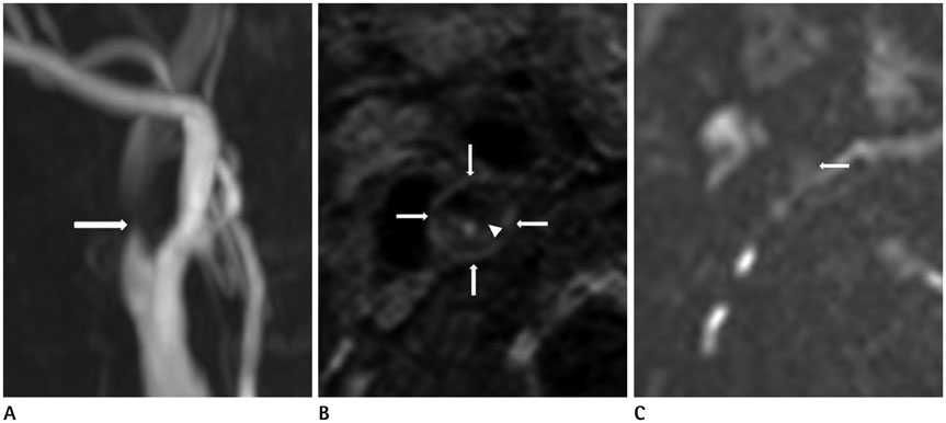
Fig. 2 A 78-year-old man with right symptomatic carotid stenosis. A. TOF MR angiography shows an eccentric plaque of the right proximal internal carotid artery (arrow). Maximal wall thickness is 5.2 mm and the longitudinal length of the plaque is 16 mm. B. Black-blood contrast-enhanced MR imaging shows large LRNC (arrows) with thin/ruptured fibrous cap (arrowhead). C. DWI with b 200 s/mm2 shows an ill-defined margin of carotid plaque due to partial volume artifact (arrow). DWI = diffusion-weighted imaging, LRNC = lipid-rich necrotic core, TOF = time of flight

Fig. 3 Contrast-noise ratio of each b-value in the DWI-identified group. DWI = diffusion-weighted imaging
Cited by 1 articles
-
The Safety of Protected Carotid Artery Stenting in Patients with Unstable Plaque on Carotid High-Resolution MR Imaging
Jae Yeong Jeong, Hyo Sung Kwak, Seung Bae Hwang, Gyung Ho Chung
J Korean Soc Radiol. 2018;78(6):380-388. doi: 10.3348/jksr.2018.78.6.380.
Reference
-
1. Smilde TJ, van Wissen S, Wollersheim H, Trip MD, Kastelein JJ, Stalenhoef AF. Effect of aggressive versus conventional lipid lowering on atherosclerosis progression in familial hypercholesterolaemia (ASAP): a prospective, randomised, double-blind trial. Lancet. 2001; 357:577–581.2. Zhao XQ, Dong L, Hatsukami T, Phan BA, Chu B, Moore A, et al. MR imaging of carotid plaque composition during lipid-lowering therapy a prospective assessment of effect and time course. JACC Cardiovasc Imaging. 2011; 4:977–986.3. Migrino RQ, Bowers M, Harmann L, Prost R, LaDisa JF Jr. Carotid plaque regression following 6-month statin therapy assessed by 3T cardiovascular magnetic resonance: comparison with ultrasound intima media thickness. J Cardiovasc Magn Reson. 2011; 13:37.4. Takaya N, Yuan C, Chu B, Saam T, Polissar NL, Jarvik GP, et al. Presence of intraplaque hemorrhage stimulates progression of carotid atherosclerotic plaques: a high-resolution magnetic resonance imaging study. Circulation. 2005; 111:2768–2775.5. Sun J, Underhill HR, Hippe DS, Xue Y, Yuan C, Hatsukami TS. Sustained acceleration in carotid atherosclerotic plaque progression with intraplaque hemorrhage: a long-term time course study. JACC Cardiovasc Imaging. 2012; 5:798–804.6. Sun J, Balu N, Hippe DS, Xue Y, Dong L, Zhao X, et al. Subclinical carotid atherosclerosis: short-term natural history of lipid-rich necrotic core--a multicenter study with MR imaging. Radiology. 2013; 268:61–68.7. Takaya N, Yuan C, Chu B, Saam T, Underhill H, Cai J, et al. Association between carotid plaque characteristics and subsequent ischemic cerebrovascular events: a prospective assessment with MRI--initial results. Stroke. 2006; 37:818–823.8. Parmar JP, Rogers WJ, Mugler JP 3rd, Baskurt E, Altes TA, Nandalur KR, et al. Magnetic resonance imaging of carotid atherosclerotic plaque in clinically suspected acute transient ischemic attack and acute ischemic stroke. Circulation. 2010; 122:2031–2038.9. Turc G, Oppenheim C, Naggara O, Eker OF, Calvet D, Lacour JC, et al. Relationships between recent intraplaque hemorrhage and stroke risk factors in patients with carotid stenosis: the HIRISC study. Arterioscler Thromb Vasc Biol. 2012; 32:492–499.10. Toussaint JF, LaMuraglia GM, Southern JF, Fuster V, Kantor HL. Magnetic resonance images lipid, fibrous, calcified, hemorrhagic, and thrombotic components of human atherosclerosis in vivo. Circulation. 1996; 94:932–938.11. Shinnar M, Fallon JT, Wehrli S, Levin M, Dalmacy D, Fayad ZA, et al. The diagnostic accuracy of ex vivo MRI for human atherosclerotic plaque characterization. Arterioscler Thromb Vasc Biol. 1999; 19:2756–2761.12. Fayad ZA, Fuster V. Characterization of atherosclerotic plaques by magnetic resonance imaging. Ann N Y Acad Sci. 2000; 902:173–186.13. Yuan C, Mitsumori LM, Ferguson MS, Polissar NL, Echelard D, Ortiz G, et al. In vivo accuracy of multispectral magnetic resonance imaging for identifying lipid-rich necrotic cores and intraplaque hemorrhage in advanced human carotid plaques. Circulation. 2001; 104:2051–2056.14. Yuan C, Kerwin WS, Ferguson MS, Polissar N, Zhang S, Cai J, et al. Contrast-enhanced high resolution MRI for atherosclerotic carotid artery tissue characterization. J Magn Reson Imaging. 2002; 15:62–67.15. Kim SE, Jeong EK, Shi XF, Morrell G, Treiman GS, Parker DL. Diffusion-weighted imaging of human carotid artery using 2D single-shot interleaved multislice inner volume diffusion-weighted echo planar imaging (2D ss-IMIV-DWEPI) at 3T: diffusion measurement in atherosclerotic plaque. J Magn Reson Imaging. 2009; 30:1068–1077.16. Kim SE, Treiman GS, Roberts JA, Jeong EK, Shi X, Hadley JR, et al. In vivo and ex vivo measurements of the mean ADC values of lipid necrotic core and hemorrhage obtained from diffusion weighted imaging in human atherosclerotic plaques. J Magn Reson Imaging. 2011; 34:1167–1175.17. Young VE, Patterson AJ, Sadat U, Bowden DJ, Graves MJ, Tang TY, et al. Diffusion-weighted magnetic resonance imaging for the detection of lipid-rich necrotic core in carotid atheroma in vivo. Neuroradiology. 2010; 52:929–936.18. Yuan C, Kerwin WS, Yarnykh VL, Cai J, Saam T, Chu B, et al. MRI of atherosclerosis in clinical trials. NMR Biomed. 2006; 19:636–654.19. Underhill HR, Yarnykh VL, Hatsukami TS, Wang J, Balu N, Hayes CE, et al. Carotid plaque morphology and composition: initial comparison between 1.5- and 3.0-T magnetic field strengths. Radiology. 2008; 248:550–560.20. Cai J, Hatsukami TS, Ferguson MS, Kerwin WS, Saam T, Chu B, et al. In vivo quantitative measurement of intact fibrous cap and lipid-rich necrotic core size in atherosclerotic carotid plaque: comparison of high-resolution, contrast-enhanced magnetic resonance imaging and histology. Circulation. 2005; 112:3437–3444.21. Saam T, Ferguson MS, Yarnykh VL, Takaya N, Xu D, Polissar NL, et al. Quantitative evaluation of carotid plaque composition by in vivo MRI. Arterioscler Thromb Vasc Biol. 2005; 25:234–239.22. Lindsay AC, Biasiolli L, Lee JM, Kylintireas I, MacIntosh BJ, Watt H, et al. Plaque features associated with increased cerebral infarction after minor stroke and TIA: a prospective, case-control, 3-T carotid artery MR imaging study. JACC Cardiovasc Imaging. 2012; 5:388–396.23. Allkemper T, Tombach B, Schwindt W, Kugel H, Schilling M, Debus O, et al. Acute and subacute intracerebral hemorrhages: comparison of MR imaging at 1.5 and 3.0 T--initial experience. Radiology. 2004; 232:874–881.24. Zhu DC, Ferguson MS, DeMarco JK. An optimized 3D inversion recovery prepared fast spoiled gradient recalled sequence for carotid plaque hemorrhage imaging at 3.0 T. Magn Reson Imaging. 2008; 26:1360–1136.25. Ota H, Yarnykh VL, Ferguson MS, Underhill HR, Demarco JK, Zhu DC, et al. Carotid intraplaque hemorrhage imaging at 3.0-T MR imaging: comparison of the diagnostic performance of three T1-weighted sequences. Radiology. 2010; 254:551–563.26. Wang J, Ferguson MS, Balu N, Yuan C, Hatsukami TS, Börnert P. Improved carotid intraplaque hemorrhage imaging using a slab-selective phase-sensitive inversion-recovery (SPI) sequence. Magn Reson Med. 2010; 64:1332–1340.27. Wasserman BA, Smith WI, Trout HH 3rd, Cannon RO 3rd, Balaban RS, Arai AE. Carotid artery atherosclerosis: in vivo morphologic characterization with gadolinium-enhanced double-oblique MR imaging initial results. Radiology. 2002; 223:566–573.28. Clarke SE, Hammond RR, Mitchell JR, Rutt BK. Quantitative assessment of carotid plaque composition using multicontrast MRI and registered histology. Magn Reson Med. 2003; 50:1199–1208.29. Qiao Y, Ronen I, Viereck J, Ruberg FL, Hamilton JA. Identification of atherosclerotic lipid deposits by diffusion-weighted imaging. Arterioscler Thromb Vasc Biol. 2007; 27:1440–1446.
- Full Text Links
-
- Actions
-
Cited
- CITED
-
- Close
- Share
-
- Similar articles
-
- Massive Cerebral Microemboli after Protected Carotid Artery Angioplasty and Stenting Using a Distal Filter Embolic Protection Device for a Vulnerable Plaque with a Lipid Rich Necrotic Core and Intraplaque Hemorrhage: A Case Report
- Findings of Angiography and Carotid Vessel Wall Imaging Associated with Post-Procedural Clinical Events after Carotid Artery Stenting
- Analysis of ¹â¸F-Fluorodeoxyglucose and ¹â¸F-Fluoride Positron Emission Tomography in Korean Stroke Patients with Carotid Atherosclerosis
- Carotid color doppler flow imaging of cerebral infarction in korea
- Multiple Carotid Artery Occlusive Diseases Treated with Staged Subclavian-carotid Artery bypass and Carotid Endarterectomy: Case Report
