Undermining and Ballooning the Proximal Part of the Left Main Coronary Artery Stent Resulting in an Iatrogenic Stent Deformation
- Affiliations
-
- 1Department of Cardiology, Cardiovascular Center, Korea University Anam Hospital, Seoul, Korea. psyche94@gmail.com
- KMID: 2151825
- DOI: http://doi.org/10.12997/jla.2015.4.2.149
Abstract
- We present a case of a 52-year-old woman with iatrogenic stent deformation occurred after deployment of the left main (LM) stent due to the unintentional undermining of the proximal part of the LM stent with subsequent balloon dilatation. We tried to crush the deformed part of the LM stent against the left coronary cusp by pushing it with a guiding catheter. The deformed stent was stabilized after stent crushing and the patient didn't have any cardiovascular events. This case highlights that stent deformation could be successfully managed by crushing the deformed part of the stent to the coronary sinus.
MeSH Terms
Figure
-
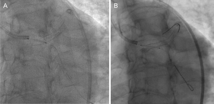
Fig. 1 (A) Coronary angiogram shows that the Choice PT wire undermined the proximal part of left main stent. (B) Deformed left main stent was hanging at the left coronary cusp.
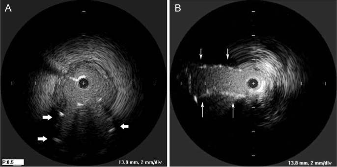
Fig. 2 (A) Intravascular ultrasound (IVUS) showed the deformed stent protruding to the left coronary cusp, (B) Follow-up IVUS showed the crushed stent struts (arrows) toward the left coronary cusp.
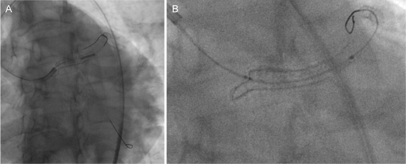
Fig. 3 (A) Crushing the deformed part against the left coronary cusp by pushing the protruded stent with the guiding catheter, (B) Coronary angiogram showed the crushed stent.

Fig. 4 (A) The three-dimensional (3D) contrast-enhanced coronary CT angiography showed a segment (4 mm in size) of the left main coronary stent protruding from the coronary artery orifice into the aorta after crushing the deformed part with the guiding catheter, (B) 3D contrast-enhanced coronary CT angiography showed the crushed stent at oblique view. Arrows indicate stent struts.
Reference
-
1. Ota H, Kitabata H, Magalhaes MA, Bui A, Kardenas K, Thomas CH, et al. Comparison of frequency and severity of longitudinal stent deformation among various drug-eluting stents: an intravascular ultrasound study. Int J Cardiol. 2014; 175:261–267.
Article2. Al-Moghairi AM, Al-Amri HS. Management of retained intervention guide-wire: a literature review. Curr Cardiol Rev. 2013; 9:260–266.
Article3. Trehan V, Mukhopadhyay S, Yusuf J, C Ramgasetty U, Mukherjee S, Arora R. Intracoronary fracture and embolization of a coronary angioplasty balloon catheter: retrieval by a simple technique. Catheter Cardiovasc Interv. 2003; 58:473–477.
Article4. Collins N, Horlick E, Dzavik V. Triple wire technique for removal of fractured angioplasty guidewire. J Invasive Cardiol. 2007; 19:E230–E234.5. Alomar ME, Michael TT, Patel VG, Altomare CG, Rangan BV, Cipher D, et al. Stent loss and retrieval during percutaneous coronary interventions: a systematic review and meta-analysis. J Invasive Cardiol. 2013; 25:637–641.6. Cha KS. Surgical retrieval of dislodged stent during transradial coronary intervention. J Invasive Cardiol. 2012; 24:E179–E181.7. Prasad A, Ilapakurti M, Ravandi A. Successful retrieval of a frayed coronary stent using the peripheral crossover technique. J Invasive Cardiol. 2011; 23:E69–E71.
- Full Text Links
-
- Actions
-
Cited
- CITED
-
- Close
- Share
-
- Similar articles
-
- Coronary Stent Fracture in a Patient with an Atrial Septal Defect and Severe Pulmonary Hypertension
- Spontaneous resolution of new coronary artery aneurysm following guideline-directed medical therapy after drug-eluting stent implantation
- Dislodgement of Two Stents in One Patient during Percutaneous Coronary Intervention
- Percutaneous Treatment of an Injured Coronary Stent Using the Looping Wire Technique
- Treatment of Stent Dislodgement Complicated by Coronary Artery Dissection using Parallel Wire Technique and Small Balloon
