Prog Med Phys.
2015 Dec;26(4):280-285. 10.14316/pmp.2015.26.4.280.
Evaluation of Fabricated Semiconductor Sensor for Verification of gamma-ray Distribution in Brachytherapy
- Affiliations
-
- 1Department of Radiation Oncology, YeonSei Cancer Center, Seoul, Korea.
- 2Department of Biomedical Engineering, Inje University, Gimhae, Korea.
- 3Department of Radiation Oncology, School of Medicine, Yonsei University, Seoul, Korea.
- 4Department of Radiation Oncology, Haeundae Back Hospital, Busan, Korea.
- 5Division of Heavy Ion Clinical Research, Korea Institute of Radiological and Medical Sciences, Seoul, Korea.
- 6Department of Radiation Oncology, Korea Institute of Radiation Oncology and Medical Physics, Seoul, Korea.
- 7Department of Radiation Oncology, Inha University Hospital, Incheon, Korea.
- 8Department of Radiation Oncology, Busan Back Hospital, Busan, Korea. physicist@paik.ac.kr
- KMID: 2151763
- DOI: http://doi.org/10.14316/pmp.2015.26.4.280
Abstract
- In radiation therapy fields, a brachytherapy is a treatment that kills lesion of cells by inserting a radioisotope that keeps emitting radiation into the body. We currently verify the consistency of radiation treatment plan and dose distribution through film/screen system (F/S system), provide therapy after checking dose. When we check dose distribution, F/S systems have radiation signal distortion because there is low resolution by penumbra depending on the condition of film developed. In this study, We fabricated a HgI2 Semiconductor radiation sensor for base study in order that we verify the real dose distribution weather it's same as plans or not in brachytherapy. Also, we attempt to evaluate the feasibility of QA system by utilizing and evaluating the sensor to bracytherapy source. As shown in the result of detected signal with various source-to-detector distance (SDD), we quantitatively verified the real range of treatment which is also equivalent to treatment plans because only the low signal estimated as scatters was measured beyond the range of treatment. And the result of experiment that we access reproducibility on the same condition of gamma-ray, we have made sure that the CV (coefficient of variation) is within 1.5 percent so we consider that the HgI2 sensor is available at QA of brachytherapy based on the result.
MeSH Terms
Figure
-
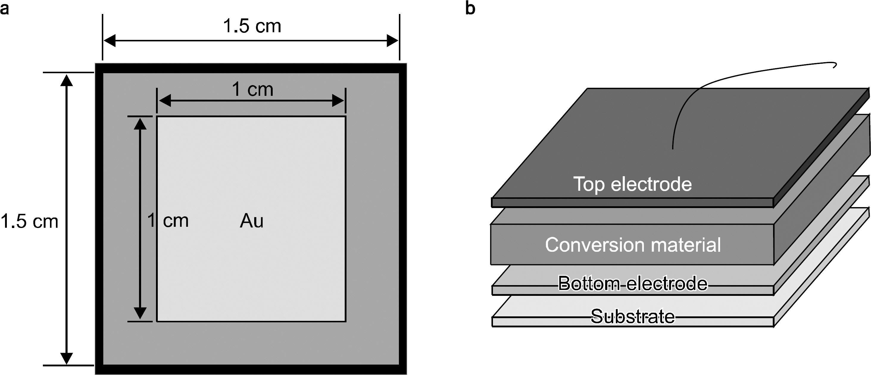
Fig. 1. Structure of material layer (a) and (b) diagram of fabricated sensors.
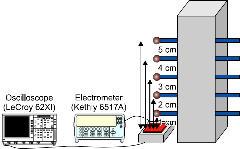
Fig. 2. Experimental setup.
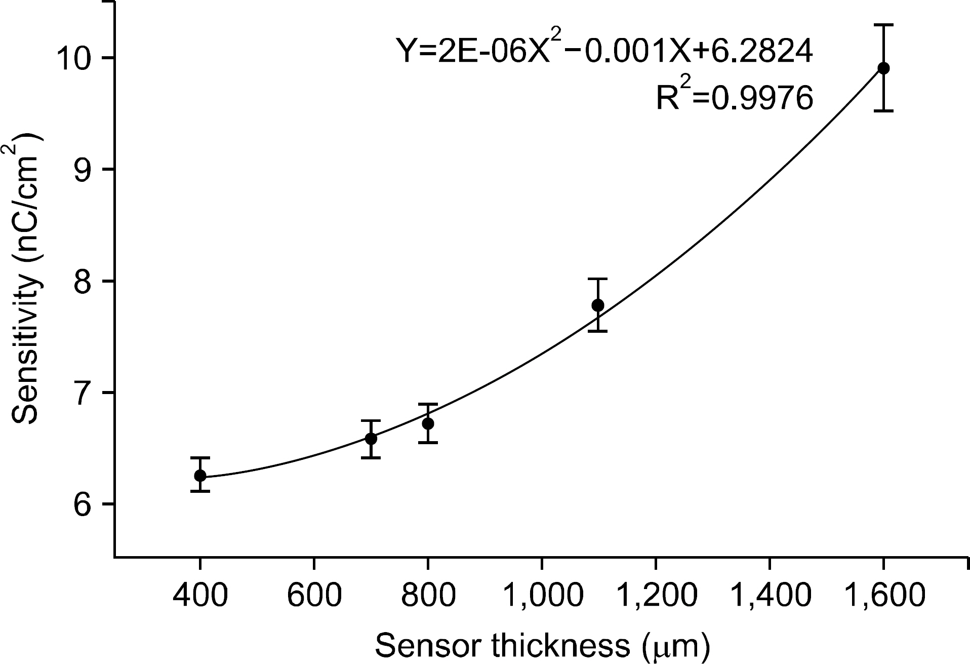
Fig. 3. Corrected signals by increased thickness respectively.
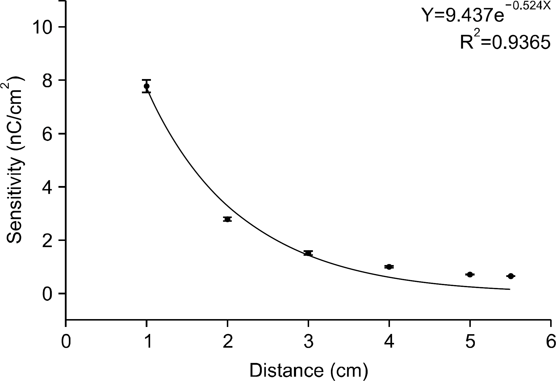
Fig. 4. Corrected signals by increased distance.
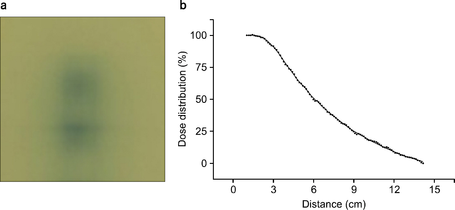
Fig. 5. Comparison of film and digital sensor.
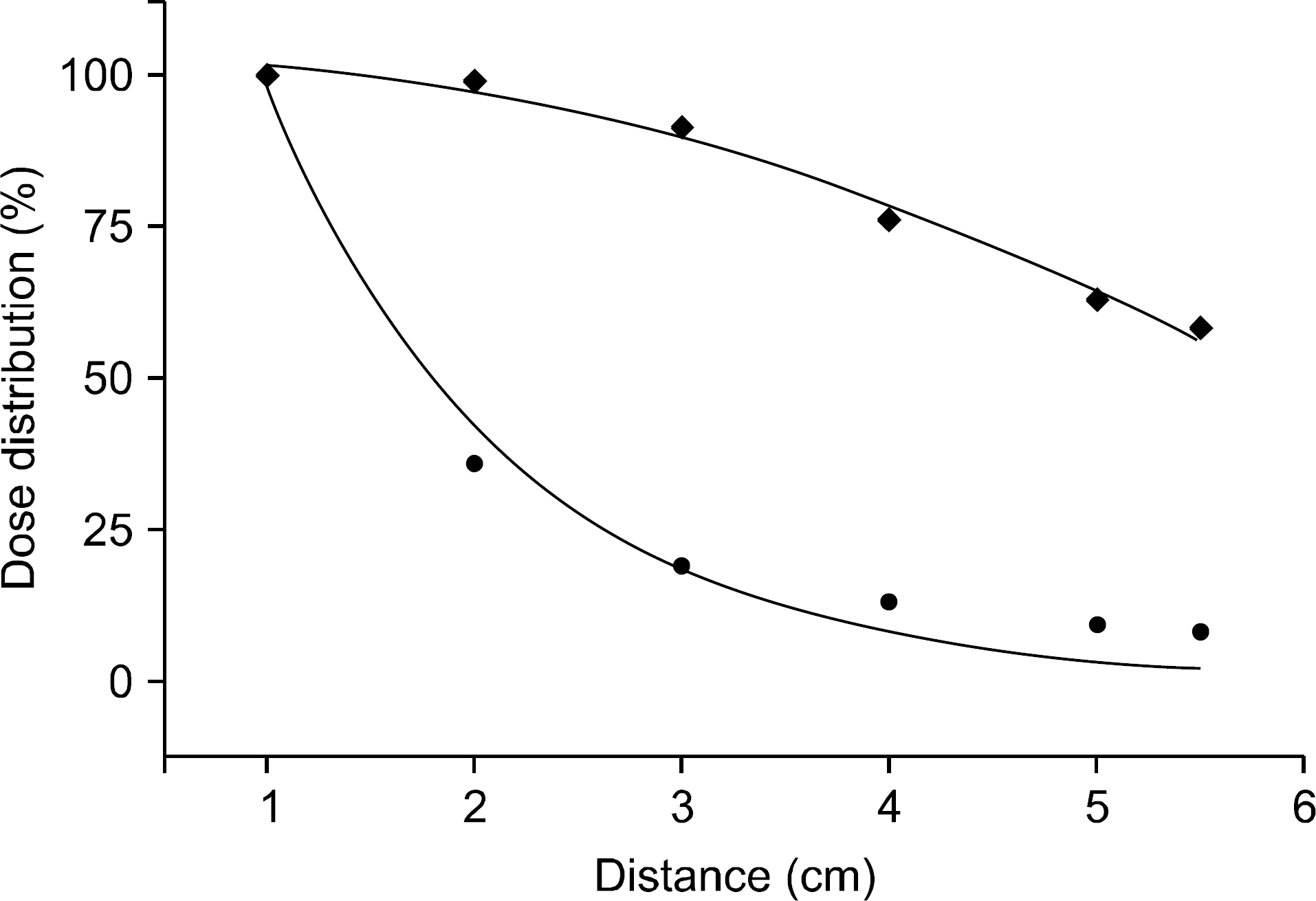
Fig. 6. Verification of γ-ray distribution by EBT film.
Reference
-
References
1. Philippe Nickers, Benedicte Thissen, Nicolas Jansen, J.M. Deneufbourg: 192Ir or 125I Prostate brachytherapy as a boost to external beam radiotherapy in locally advanced prostatic cancer: A dosimetric point of view. Radiotherapy and oncology. 78(1):47–52. 2006.2. Giwon Jang, Jungwook Shin, Kyungmin Oh, et al. The Feasibility Study of photoconductor materials for the use of adosimeter in Radiotherapy. The Korean Society of Radiology. 7(1):81–84. 2013.3. Bosun Kang, et al. Development of image quality evaluation program for diagnostic radiography. The Korean Society of Radiography. 2(2):5–10. 2008.4. Qihua Zhao. Larry E. Antonuk, Youcef EI-Mohri, et al: Performance evaluation of polycrystalline HgI2 photoconductors for radiation therapy imaging. Med Phys. 37(6):2738–2748. 2010.5. Youngbin Kim, Minseok Yun, Minwoo Kim, et al. Development of Radiation Image Sensor using Hetero-junction. The Korean Society of Radiography. 3(3):27–35. 2009.6. Kyotae Kim, Sangsik Kang, Sicheul Noh, et al. Absorbed Spectrum Comparison of Lead and Tungsten in Continuous X-ray Energy using Monte Carlo Simulation. The Korean Society of Radiography. 6(6):483–487. 2012.7. Sangsik Kang, Kyotae Kim, Sicheol Noh, et al. The Study on Design of Customized Radiation Protective Layer for Medical Radiation Dose Reduction. The Korean Society of Radiography. 8(6):333–338. 2014.8. J. H. Hubbell, S. M. Seltzer, et al: Tables of X-ray Mass Attenuation Coefficients and Mass Energy-Absorption Coefficients. National Institute of Standards and Technology (. 1999.9. Sukhee Jung, Yoonsuk Kim, Youngbin Kim, et al. The study of PbO's sintering effect for high efficiency x-ray detection sensor. The Korean Society of Radiography. 3(3):37–40. 2009.10. Minseok Yun, Sungho Cho, Kyungmin Oh, et al. X-ray properties measurement of Flat panel Digital X-ray gas detector. The Korean Society of Radiography. 3(1):15–19. 2009.11. Luiz Antonio R, da Rosa: Reproducibility study of TLC-100 micro-cubes at radiothrapy dose level. Applied Radiation and Isotopes. 50:573–577. 1999.
- Full Text Links
-
- Actions
-
Cited
- CITED
-
- Close
- Share
-
- Similar articles
-
- Options in Intracoronary Radiation Therapy
- Development of Dosimetric Verification System for Patient-Specific Quality Assurance of High-Dose-Rate Brachytherapy
- Development of a Stereotactic Device for Gamma Knife Irradiation of Small Animals
- Development of Two-dimensional Prompt-gamma Measurement System for Verification of Proton Dose Distribution
- Three-dimensional dose reconstruction-based pretreatment dosimetric verification in volumetric modulated arc therapy for prostate cancer
