Dedifferentiated Retroperitoneal Liposarcoma Presenting as Right Inguinal Hernia: A Case Report
- Affiliations
-
- 1Department of Radiology, Uijeongbu St. Mary's Hospital, College of Medicine, The Catholic University of Korea, Uijeongbu, Korea. ymiku@catholic.ac.kr
- 2Department of Radiology, Seoul St. Mary's Hospital, College of Medicine, The Catholic University of Korea, Seoul, Korea.
- KMID: 2150465
- DOI: http://doi.org/10.3348/jksr.2016.74.1.66
Abstract
- Retroperitoneal liposarcomas usually present as painless, slow-growing abdominal masses. When masses grow large enough to compress surrounding structures, symptoms may occur. Retroperitoneal liposarcoma clinically manifesting as inguinal hernia is a very rare entity; only 11 cases have been reported. Herein, we present radiographic features of a 37-year-old male with a painless palpable mass in the right groin that was identified as dedifferentiated retroperitoneal liposarcoma herniated through the right inguinal canal.
MeSH Terms
Figure
-
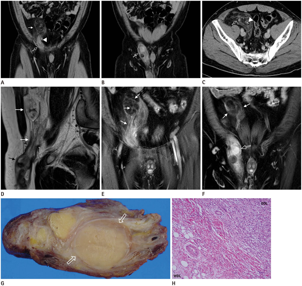
Fig. 1 A 37-year-old male with a retroperitoneal dedifferentiated liposarcoma (DDL) extending into the right inguinal canal. A, B. Contrast-enhanced computed tomography (CT) with coronal reformatted images shows a large mass with a fatty component in the retro-peritoneum (thin arrows) extending into the right inguinal canal with a solid enhancing component (open arrow). Note the medial displacement of the inferior epigastric vessels (arrowhead). C. Axial contrast-enhanced CT demonstrates several ill-defined strands and a soft-tissue nodule with homogenous enhancement (open arrow) within the mass. Note the mass adjacent to and extending along the testicular vessels (arrowhead). D. Sagittal T2-weighted image of the retroperitoneal tumor extending into the right inguinal canal. The retroperitoneal component of the mass shows high signal intensity consistent with mature fatty tissue (white arrows), while the inguinal canal component shows intermediate signal intensity consistent with tumor tissue (black arrow). E, F. Coronal gadolinium-pentetic-acid-enhanced T1-weighted images demonstrate multiple tenuous septa (arrows) and a soft-tissue nodule with marked enhancement (open arrow) within the mass. G. On cross-section, the surgical specimen taken from the right inguinal canal is solid, with discrete intratumoral nodules of varying size and colors ranging from yellow to yellow-tan admixed with firm tan-gray areas corresponding to dedifferentiated foci (open arrows). H. The tumor shows an abrupt transition between the components of well-differentiated liposarcoma and DDL (hematoxylin & eosin, × 100).
Reference
-
1. Hong SH, Kim KA, Woo OH, Park CM, Kim CH, Kim MJ, et al. Dedifferentiated liposarcoma of retroperitoneum: spectrum of imaging findings in 15 patients. Clin Imaging. 2010; 34:203–210.2. Mizuno Y, Sumi Y, Nachi S, Ito Y, Marui T, Saji S, et al. A case of a large retroperitoneal liposarcoma presenting as an incarcerated inguinal hernia. Hernia. 2006; 10:439–442.3. McKinley SK, Abreu N, Patalas E, Chang A. Large Retroperitoneal Liposarcoma Diagnosed upon Radiological Evaluation of Mild Right-Sided Inguinal Hernia. Case Rep Radiol. 2013; 2013:187957.4. Vagnoni V, Brunocilla E, Schiavina R, Borghesi M, Passaretti G, Gentile G, et al. Inguinal canal tumors of adulthood. Anticancer Res. 2013; 33:2361–2368.5. Lahat G, Anaya DA, Wang X, Tuvin D, Lev D, Pollock RE. Resectable well-differentiated versus dedifferentiated liposarcomas: two different diseases possibly requiring different treatment approaches. Ann Surg Oncol. 2008; 15:1585–1593.6. Tateishi U, Hasegawa T, Beppu Y, Satake M, Moriyama N. Primary dedifferentiated liposarcoma of the retroperitoneum. Prognostic significance of computed tomography and magnetic resonance imaging features. J Comput Assist Tomogr. 2003; 27:799–780.7. Song T, Shen J, Liang BL, Mai WW, Li Y, Guo HC. Retroperitoneal liposarcoma: MR characteristics and pathological correlative analysis. Abdom Imaging. 2007; 32:668–674.8. Nishino M, Hayakawa K, Minami M, Yamamoto A, Ueda H, Takasu K. Primary retroperitoneal neoplasms: CT and MR imaging findings with anatomic and pathologic diagnostic clues. Radiographics. 2003; 23:45–57.9. Aikawa H, Tanoue S, Okino Y, Tomonari K, Miyake H. Pelvic extension of retroperitoneal fluid: analysis in vivo. AJR Am J Roentgenol. 1998; 171:671–677.10. Aguirre DA, Santosa AC, Casola G, Sirlin CB. Abdominal wall hernias: imaging features, complications, and diagnostic pitfalls at multi-detector row CT. Radiographics. 2005; 25:1501–1520.
- Full Text Links
-
- Actions
-
Cited
- CITED
-
- Close
- Share
-
- Similar articles
-
- A Case of Dedifferentiated LiposarcomaThat Developed in the Dermis
- Dedifferentiated Liposarcoma of the Retroperitoneum: A case report
- Dedifferentiated Liposarcoma with a Peculiar Whorling Pattern: A Case Report
- Dedifferentiated Liposarcoma in the Thigh: Case Report
- Imaging Findings of Scrotal Liposarcoma: A Case Report
