J Korean Ophthalmol Soc.
2015 Jul;56(7):1122-1126. 10.3341/jkos.2015.56.7.1122.
Uveitis in Both Eyes Associated with Sweet's Syndrome
- Affiliations
-
- 1Department of Ophthalmology, Wonkwang University Hospital, Wonkwang University School of Medicine, Iksan, Korea. cuchoi77@hanmail.net
- KMID: 2148815
- DOI: http://doi.org/10.3341/jkos.2015.56.7.1122
Abstract
- PURPOSE
To report a case of uveitis in both eyes caused by Sweet's syndrome.
CASE SUMMARY
A 66-year-old male presented with decreased visual acuity in his left eye. Three years prior he was diagnosed with Sweet's syndrome, with symptoms such as chill, fever and, maculopapular rash on the chest. At initial physical examination, he had 3 or 4+ inflammatory cells and flare in the anterior chambers of both eyes, as well as hypopyon in his left eye. Under the suspicion of uveitis caused by Sweet's syndrome, he was rescribed an IV steroid injection and topical steroid agent. Three days later, his visual acuity improved to 0.3 in the right eye and 0.2 in the left eye. Hypopyon in the left eye disappeared and inflammatory cells decreased to 1~2+. He showed signs of recurrence in both eyes after 5 months and was treated with posterior subtenon triamcinolone injection in each eye. The patient showed no signs of recurrence for 10 months after injection.
CONCLUSIONS
We report a case of uveitis caused by Sweet's syndrome treated with a steroid agent resulting in good prognosis. To the best of our knowledge, this is first case of uveitis caused by Sweet's syndrome reported in Korea.
MeSH Terms
Figure
-

Figure 1. Ill-defined plaques and papules on the trunk (circles).
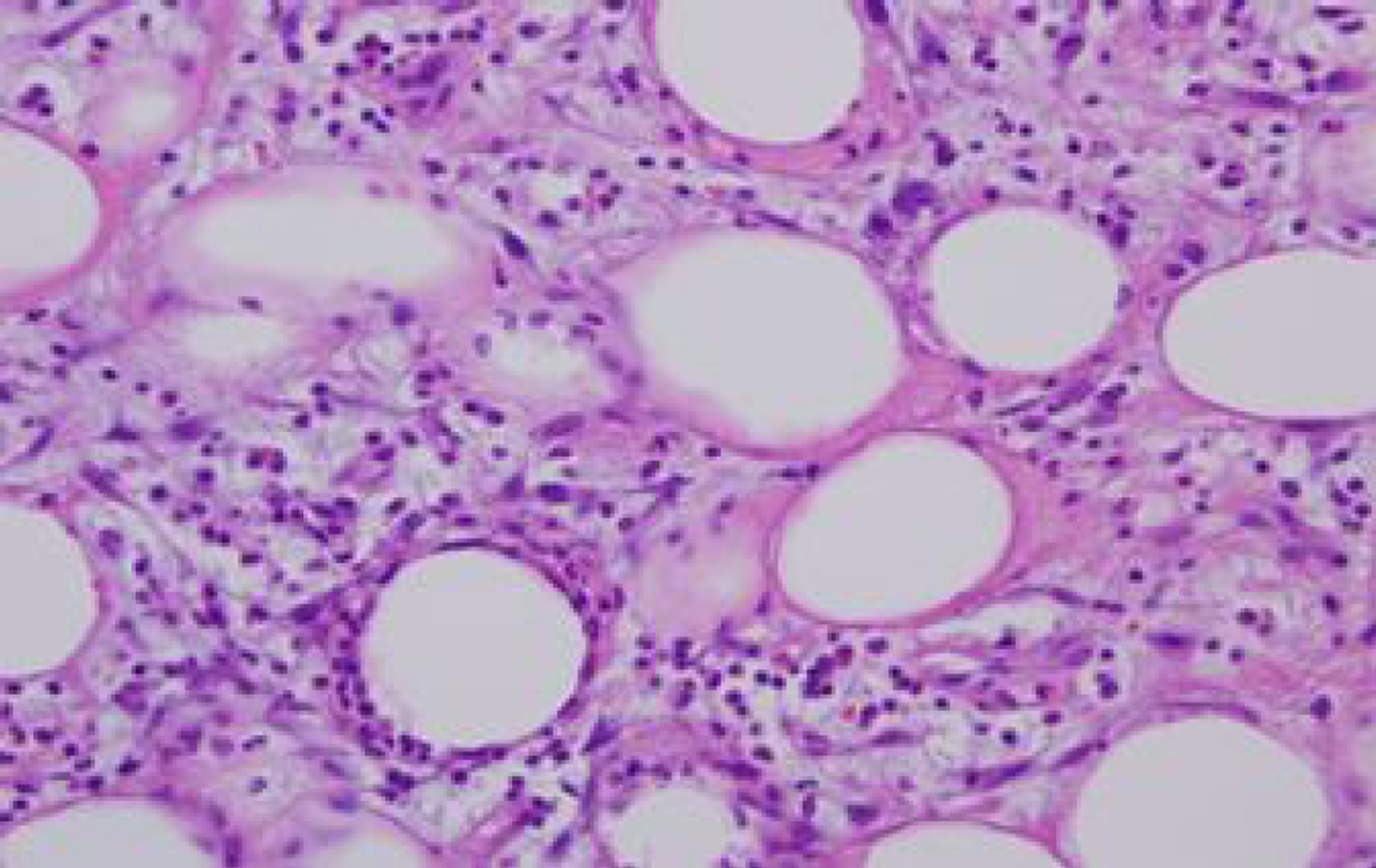
Figure 2. Histopathology of the skin demonstrating mixed sep-tal and lobular panniculitis with many neutrophilic infiltration (hematoxylin-eosin, magnification 100x).
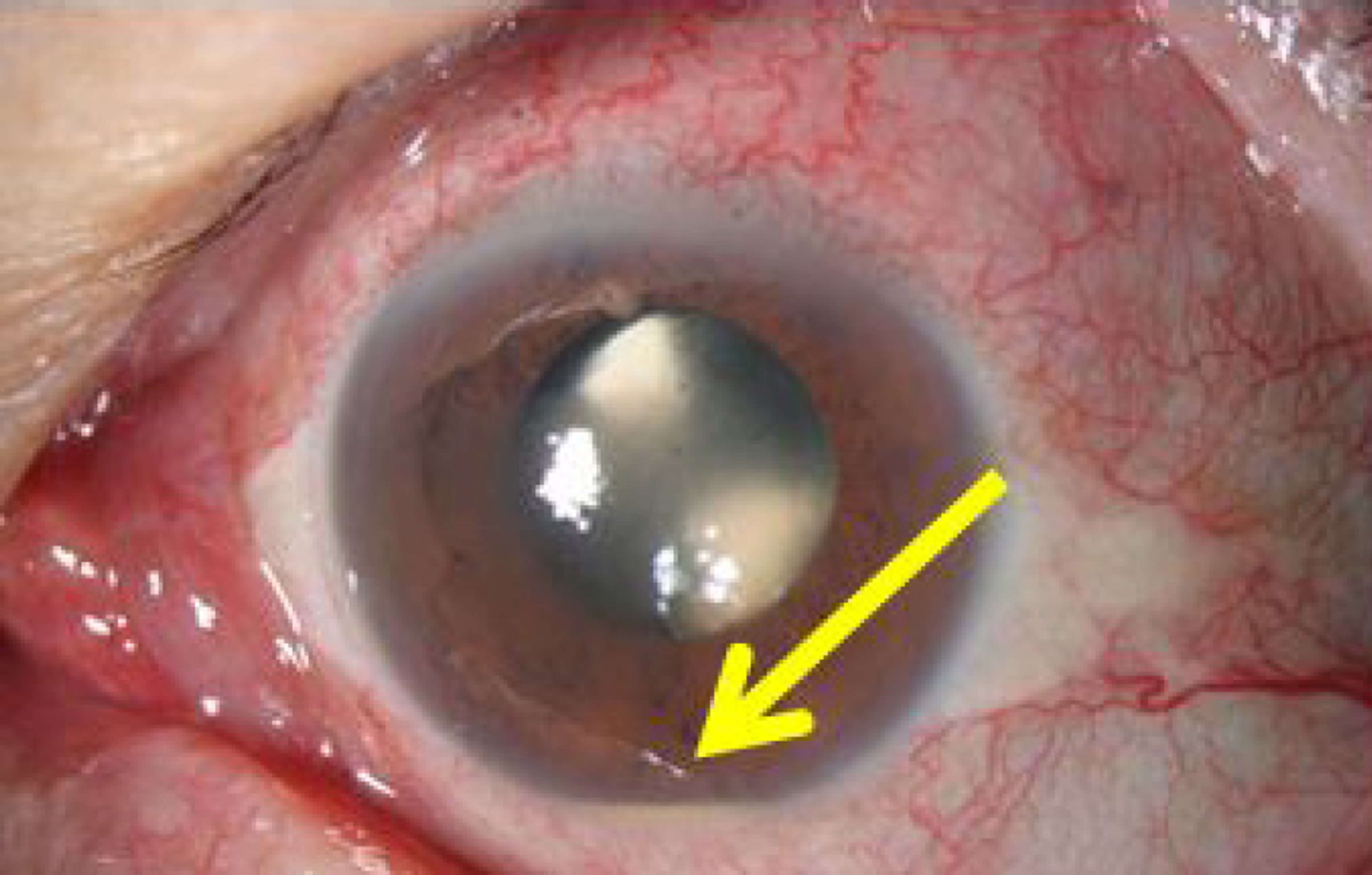
Figure 3. Hypopion in the left eye at the initial outpatient visit (arrow).

Figure 4. B scan showing inflammatory cells in the vitreous cavity demonstrating posterior uveitis. (A) Right eye. (B) Left eye.

Figure 5. Fluororescein angiography showing optic disc hyperfluorescence at the latent period. (A) Right eye. (B) Left eye.
Reference
-
References
1. Sweet RD. An acute febrile neutrophilic dermatosis. Br J Dermatol. 1964; 76:349–56.2. Cohen PR. Sweet’s syndrome: a comprehensive review of an acute febrile neutrophilic dermatosis. Orphanet J Rare Dis. 2007; 2:34.
Article3. Gottlieb CC, Mishra A, Belliveau D, et al. Ocular involvement in acute febrile neutrophilic dermatosis (Sweet syndrome): new cas-es and review of the literature. Surv Opthalmol. 2008; 53:219–26.
Article4. Limdiwala PG, Parikh SJ, Shah JS. Sweet’s syndrome. Indian J Dent Res. 2014; 25:401–5.
Article5. Michel G, Lhermitte B, Cribier B, et al. Sweet syndrome present-ing as resistant conjunctivitis. Cornea. 2008; 27:1189–90.
Article6. O’Brien MC. Sweet’s syndrome. J Emerg Med. 2005; 29:341–2.
Article7. Femiano F, Gombos F, Scully C. Sweet’s syndrome: recurrent oral ulceration, pyrexia, thrombophlebitis, and cutaneuous lesions. Oral Surg Oral Med Oral Pathol Oral Radiol Endod. 2003; 95:324–7.8. Jung HT, Kim SK, Park KW, et al. A case of Sweet syndrome in-volving the central nervous system. Korean J Med. 2008; 75:463–6.9. Ko BG, Lee HG, Lee HS, et al. A case of Sweet’s syndrome with pulmonary infiltration and pleural effusion in myelodysplastic syndrome. Korean J Med. 2011; 80:216–20.10. von den Driesch P. Sweet’s syndrome (acute febrile neutrophilic dermatosis). J Am Acad Dermatol. 1994; 31:535–56. quiz. 557–60.
Article11. Medenblik-Frysch S, von den Driesch P, Jonas JB, Meythaler FH. Ocular complications in Sweet’s syndrome. Am J Ophthalmol. 1992; 114:230–1.
Article12. Fourman S. Inflammatory glaucoma associated with Sweet’s syndrome. Am J Opthalmol. 1988; 105:691–2.
Article13. Mazpakis E, Kalikaki A, Stathopoulos E, et al. Acute febrile neu-trophilic dermatosis (Sweet’s syndrome) with erythema nodosum and anterior scleritis. Int J Dermatol. 2005; 44:1051–3.14. Song WK, Bang D, Choi YJ, et al. Sudden visual loss due to occlu-sive venous vasculitis associated with Sweet syndrome. Arch Dermatol. 2009; 145:216–8.
Article15. Sato M, Kawamura T, Hase S, et al. A case of bilateral retinal vas-culitis associated with Sweet syndrome. Retina. 2005; 25:800–2.
Article16. Lobo AM, Stacy R, Cestari D, et al. Optic nerve involvement with panuveitis in Sweet syndrome. Ocul immunol inflamm. 2011; 19:167–70.
Article17. Matsumiya W, Kusuhara S, Yamada Y, et al. Sweet’s syndrome with panuveitis resembling Behçet’s disease. Jpn J Ophthalmol. 2012; 56:268–72.
Article18. Cohen PR, Kurzrock R. Sweet’s syndrome: a neutrophilic dermatosis classically associated with acute onset and fever. Clin Dermatol. 2000; 18:265–82.
Article19. Su WP, Liu HN. Diagnostic criteria for Sweet's syndrome. Cutis. 1986; 37:167–74.20. Gunawardena DA, Gunawardena KA, Ratnayaka RM, Vasanthana-than NS. The clinical spectrum of Sweet’s syndrome (acute febrile neutrophilic dermatosis)- a report of eighteen cases. Br J Dermatol. 1975; 92:363–73.
- Full Text Links
-
- Actions
-
Cited
- CITED
-
- Close
- Share
-
- Similar articles
-
- Sweet's Syndrome Associated with Bacterial Meningitis
- Neuro-Sweet's Syndrome with Central Nervous System Involvement
- Sweet's Syndrome with Myelodysplastic Syndrome Progressing to Acute Myelogenous Leukemia
- Sweet's Syndrome Associated with Acute Erythema Nodosum
- Postoperative outcome of trans pars pklana vitrectomy in the treatment of posterior and intermediate uveitis
