J Korean Ophthalmol Soc.
2015 Dec;56(12):1969-1973. 10.3341/jkos.2015.56.12.1969.
Radius-Maumenee Syndrome Presenting with Ocular Pain and Conjunctival Injection: A Case Report
- Affiliations
-
- 1Department of Ophthalmology, Seoul National University College of Medicine, Seoul, Korea. eye@snubh.org
- 2Department of Ophthalmology, Seoul National University Bundang Hospital, Seongnam, Korea.
- KMID: 2148763
- DOI: http://doi.org/10.3341/jkos.2015.56.12.1969
Abstract
- PURPOSE
Radius-Maumenee syndrome (RMS) is characterized by idiopathic dilated episcleral vessels usually associated with glaucoma. The authors report a case of a 32-year-old Korean male with dilation of the episcleral vessels and glaucoma in both eyes.
CASE SUMMARY
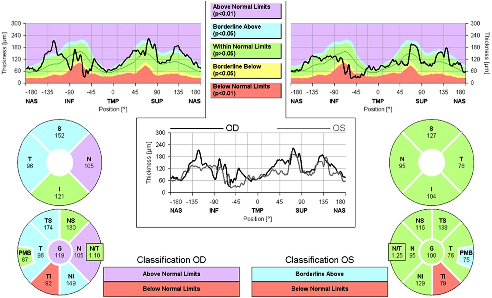
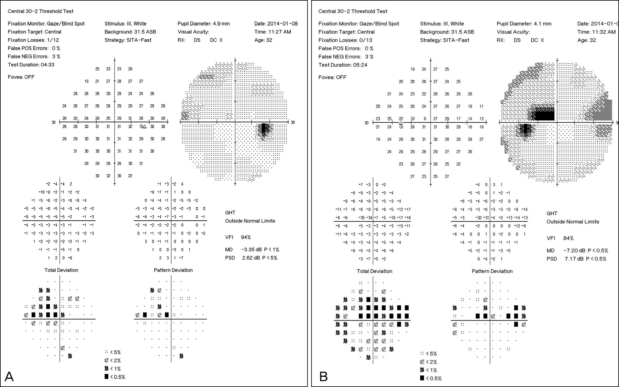
A 32-year-old Korean male presented with conjunctival injection and chronic pulsatile ocular pain in both eyes for 11 years. His best corrected visual acuity was 20/20 in both eyes. Slit lamp biomicroscopy showed episcleral venous engorgement in both eyes. Fundus photographs revealed superotemporal and inferotemporal retinal nerve fiber layer defects and Humphrey visual field testing showed superior nasal steps and paracentral scotomas in both eyes. He suffered from chronic eye pain in both eyes although he had applied glaucoma medication and his symptoms had worsened during the past year. Brain magnetic resonance imaging (MRI) and magnetic resonance angiography (MRA) revealed no evidence of carotid cavernous fistula or other orbital lesions. Due to the presence of episcleral venous engorgement, glaucoma and negative tests for other possible diseases, he was diagnosed with RMS.
CONCLUSIONS
RMS is an idiopathic disease with episcleral vessel dilation and frequently associated with glaucoma. Its diagnosis is confirmed by eliminating other possible causes of episcleral venous engorgement.
MeSH Terms
Figure
Reference
-
References
1. Radius RL, Maumenee AE. Dilated episcleral vessels and open-an-gle glaucoma. Am J Ophthalmol. 1978; 86:31–5.
Article2. Acaroglu G, Eranil S, Ozdamar Y. . Idiopathic episcleral ve-nous engorgement. Clin Exp Optom. 2009; 92:507–10.
Article3. Stock RA, Fernandes NL, Pastro NL. . Idiopathic dilated epis-cleral vessels (Radius-Maumenee syndrome): case report. Arq Bras Oftalmol. 2013; 76:45–7.
Article4. Parikh RS, Desai S, Kothari K. Dilated episcleral veins with secon-dary open angle glaucoma. Indian J Ophthalmol. 2011; 59:153–5.
Article5. Grieshaber MC, Dubler B, Knodel C. . Retrobulbar blood flow in idiopathic dilated episcleral veins and glaucoma. Klin Monbl Augenheilkd. 2007; 224:320–3.6. Lee JJ, Yap EY. Optociliary shunt vessels in diabetes mellitus. Singapore Med J. 2004; 45:166–9.7. Lämmer R. Secondary open angle glaucoma with idiopathic epis-cleral venous pressure (Radius-Maumenee syndrome). Sinus-oto-my as operative procedure of choice. Ophthalmologe. 2007; 104:515–6.
- Full Text Links
- Actions
-
Cited
- CITED
-
- Close
- Share
- Similar articles
-
- Ocular Inflammation with Use of Oral Bisphosphonates
- Toxic Epidermal Necrolysis with Ocular Involvement Following Vaccination for Hemorrhagic Fever with Renal Syndrome
- A Case of Ocular Benign Lymphoid Hyperplasia Treated with Bevacizumab Injection
- A Case of Neovascular Glaucoma Secondary to Ocular Ischemic Syndrome in a Patient with Moyamoya Disease
- A Case of SUNCT Syndrome which Showed Marked Improvement with Carbamazepine and Discussion on Nosologic Aspect of SUNCT Syndrome


