A Case of Neovascular Glaucoma Secondary to Ocular Ischemic Syndrome in a Patient with Moyamoya Disease
- Affiliations
-
- 1Department of Ophthalmology, Pusan National University School of Medicine, Busan, Korea. alertlee@hanmail.net
Abstract
- PURPOSE
To report a case of neovascular glaucoma secondary to ocular ischemic syndrome in a patient with moyamoya disease, who was successfully treated with trabeculectomy.
CASE SUMMARY
A 45-year-old woman suffered from slowly decreased vision in the right eye 3 months previously. Ocular pain with conjunctival injection of the right eye and headache developed 2 months earlier. She was diagnosed with moyamoya disease and had an encephaloduroarteriosynangiosis at the neurosurgery. The patient complained of persistent conjunctival injection and decreased vision of the right eye after surgery. At the initial visit, best corrected visual acuity (BCVA) of the right eye was 0.1 and intraocular pressure (IOP) was 42 mm Hg. Slit lamp examination revealed neovascularization of the iris and gonioscopy showed a 360degrees peripheral anterior synechiae. Fluorescein angiography demonstrated prolonged arteriovenous transit time in the right eye. On the electroretinogram, the amplitude of both a and b waves decreased in the right eye more than in the left eye. On the magnetic resonance angiography, narrowing of the right internal carotid artery was observed. The patient was diagnosed with neovascular glaucoma due to ocular ischemic syndrome caused by moyamoya disease. Panretinal photocoaguration, intravitreal bevacizumab injection and trabeculectomy with mitomycin-C soaking was performed in the right eye. At 8 months after surgery, BCVA of the right eye was 0.1, IOP was 17 mm Hg without antiglaucoma medication and bleb was maintained in good condition.
CONCLUSIONS
The patient's results indicate that neovascular glaucoma can occur secondary to ocular ischemic syndrome caused by moyamoya disease.
MeSH Terms
-
Antibodies, Monoclonal, Humanized
Blister
Carotid Artery, Internal
Eye
Female
Fluorescein Angiography
Glaucoma, Neovascular
Gonioscopy
Headache
Humans
Intraocular Pressure
Iris
Magnetic Resonance Angiography
Middle Aged
Mitomycin
Moyamoya Disease
Neurosurgery
Trabeculectomy
Vision, Ocular
Visual Acuity
Bevacizumab
Antibodies, Monoclonal, Humanized
Mitomycin
Figure
-

Figure 1 Magnetic resonance angiogram (MRA). (A) Narrowing of right internal carotid artery (white arrow). (B) Collateral vessels on the base of brain (white arrow).
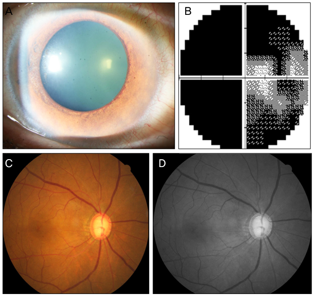
Figure 2 (A) Slit lamp photograph of the right eye shows neovascularization of the iris and dilated pupil of the right eye at the initial visit. (B) Visual field examination of the right eye reveals remaining of central and temporal islands. (C) Color fundus photograph shows dot retinal hemorrhages at the mid-periphery of the retina, narrowed retinal artery, and dilated retinal vein. Vertical cup-disc ratio was 0.9. (D) Red-free fundus photograph shows superior and inferior retinal nerve fiber layer defects.
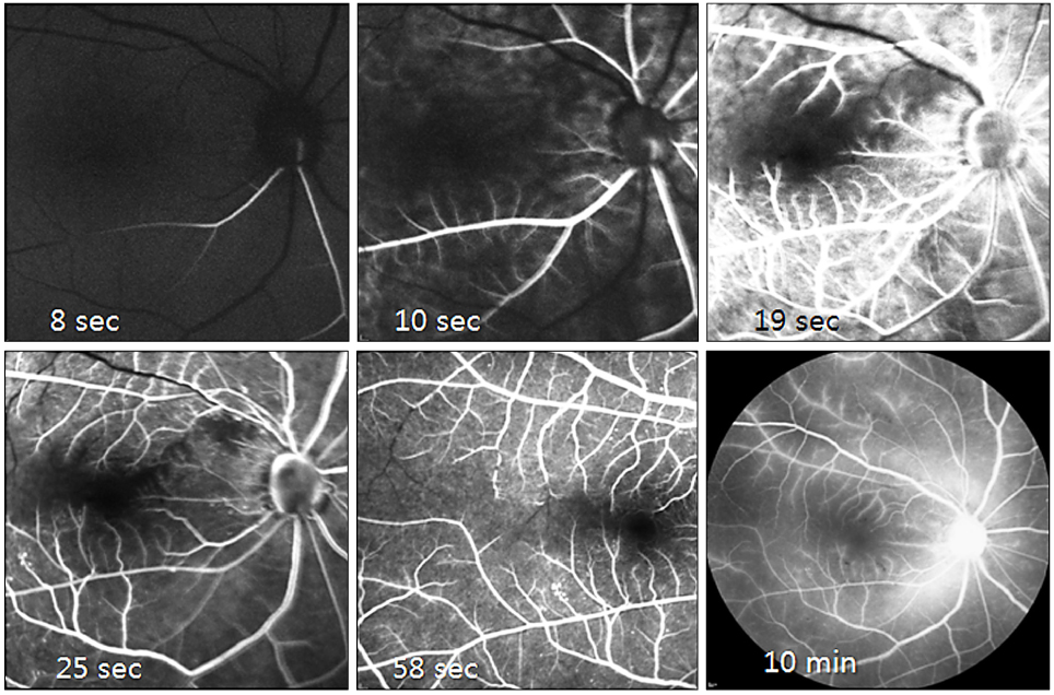
Figure 3 Fluorescent angiograph of the right eye. Choroidal filling is delayed, patchy choroidal filling is shown and leading edge of dye is seen. Arteriovenous transit time is delayed to about 50 second and venous filling time is about 58 second. Retinal capillary nonperfusion and microaneurysms are found. On the late stage, there are staining of the vessels, especially on the artery and arteriole.
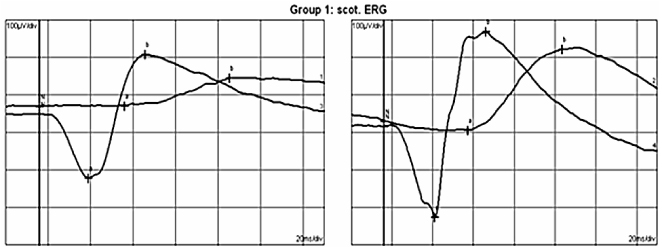
Figure 4 Electroretinogram shows a wave and b wave amplitudes of the right eye decreased more than those of the left eye.
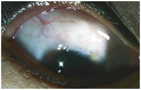
Figure 5 Slit lamp photography of the right eye shows well elevated bleb with medium height, 4 clock hour horizontal extent and mild vascularity at 8 month after trabeculectomy with mitomycin C.
Reference
-
1. Scott RM, Smith ER. Moyamoya disease and moyamoya syndrome. N Engl J Med. 2009. 360:1226–1237.2. Kuroda S, Houkin K. Moyamoya disease: current concepts and future perspectives. Lancet Neurol. 2008. 7:1056–1066.3. Noda S, Hayasaka S, Setogawa T, Matsumoto S. Ocular symptoms of moyamoya disease. Am J Ophthalmol. 1987. 103:812–816.4. Kim JH, Kim JY. A case of ophthalmic artery occlusion in moyamoya disease. J Korean Ophthalmol Soc. 2007. 48:849–853.5. Kim YS, Lee KS, Kim SH, Byun YJ. Moyamoya disease with characteristic funds findings of retinal vascular insufficiency. J Korean Ophthalmol Soc. 1998. 39:2477–2483.6. Yim HB, Mo YH, Koo HM, Chung SK. A case of moyamoya disease with right homonymous hemianopsia. J Korean Ophthalmol Soc. 1992. 33:194–198.7. Cho A, Kim SY. A case of moyamoya disease initially presenting as anterior ischemic optic neuropathy. J Korean Ophthalmol Soc. 2011. 52:887–892.8. Zhou B, Ye P, Wei S. Preliminary clinical analysis of neovascular glaucoma secondary to carotid artery disease. Clin Exp Optom. 2011. 94:207–211.9. Takeuchi K, Shimizu K. Hypoplasia of the bilateral internal carotid arteries. Brain Nerve. 1957. 9:37–43.10. Suzuki J, Takaku A. Cerebrovascular "moyamoya" disease. Disease showing abnormal net-like vessels in base of brain. Arch Neurol. 1969. 20:288–299.11. Suzuki J, Kodama N. Moyamoya disease--a review. Stroke. 1983. 14:104–109.12. Scott RM, Smith JL, Robertson RL, et al. Long-term outcome in children with moyamoya syndrome after cranial revascularization by pial synangiosis. J Neurosurg. 2004. 100:2 Suppl Pediatrics. 142–149.13. Jea A, Smith ER, Robertson R, Scott RM. Moyamoya syndrome associated with Down syndrome: outcome after surgical revascularization. Pediatrics. 2005. 116:e694–e701.14. Cho HJ, Lim KO, Hwang YH, Hwang JU. Moyamoya disease initially presenting transient visual loss. J Korean Ophthalmol Soc. 2012. 53:353–356.15. Lee YW, Lee SN. A case of bilateral upgaze palsy associated with unilateral midbrain hemorrhage in moyamoya disease. J Korean Ophthalmol Soc. 2004. 45:1772–1776.16. Chen CS, Lee AW, Kelman S, Wityk R. Anterior ischemic optic neuropathy in moyamoya disease: a first case report. Eur J Neurol. 2007. 14:823–825.17. Harissi-Dagher M, Sebag M, Dagher JH, Moumdjian R. Chorioretinal atrophy in a patient with moyamoya disease. Case report. J Neurosurg. 2004. 101:843–845.18. Chace R, Hedges TR 3rd. Retinal artery occlusion due to moyamoya disease. J Clin Neuroophthalmol. 1984. 4:31–34.19. Brown GC, Magargal LE. The ocular ischemic syndrome. Clinical, fluorescein angiographic and carotid angiographic features. Int Ophthalmol. 1988. 11:239–251.20. Mun SJ, Lee KH, Lee DU, Cho NC. Clinical features of ophthalmic artery hypoperfusion. J Korean Ophthalmol Soc. 2007. 48:297–302.21. Kahn M, Green WR, Knox DL, Miller NR. Ocular features of carotid occlusive disease. Retina. 1986. 6:239–252.22. Brown GC, Magargal LE, Simeone FA, et al. Arterial obstruction and ocular neovascularization. Ophthalmology. 1982. 89:139–146.23. Coleman K, Fitzgerald D, Eustace P, Bouchier-Hayes D. Electroretinography, retinal ischaemia and carotid artery disease. Eur J Vasc Surg. 1990. 4:569–573.24. Brown GC, Magargal LE, Sergott R. Acute obstruction of the retinal and choroidal circulations. Ophthalmology. 1986. 93:1373–1382.
- Full Text Links
-
- Actions
-
Cited
- CITED
-
- Close
- Share
-
- Similar articles
-
- A Case of Ocular Ischemic Syndrome Associated with Multiple Branch Retinal Artery Occlusion and Neovascular Glaucoma
- A Case of Ocular Ischemic Syndrome with Neovascular Glaucoma
- A Case of Ocular Ischemic Syndrome Associated with Internal Carotid Artery Obstruction with Atheroma Diagnosed by Doppler Ultrasonography
- A Case of Ocular Ischemic Syndrome Associated with Right Internal Carotid Artery Obstruction
- Bilateral Neovascular Glaucoma Attributable to a Parotid Pleomorphic Adenoma
