Clin Nutr Res.
2015 Jul;4(3):137-152. 10.7762/cnr.2015.4.3.137.
Nutritional Management of Kidney Stones (Nephrolithiasis)
- Affiliations
-
- 1Department of Nephrology, Harvard Vanguard Medical Associate, Boston, MA 02115, USA. hhan1@comcast.net, haewookhan@gmail.com
- 2Harvard Vanguard Medical Associate, Clinical Instructor at Harvard Medical School, Boston, MA 02115, USA.
- 3Harvard Vanguard Medical Associates; Brigham and Women's Hospital, Boston, MA 02115, USA.
- 4Tufts University Friedman School of Nutrition and School of Medicine, Boston, MA 02111, USA.
- KMID: 2148623
- DOI: http://doi.org/10.7762/cnr.2015.4.3.137
Abstract
- The incidence of kidney stones is common in the United States and treatments for them are very costly. This review article provides information about epidemiology, mechanism, diagnosis, and pathophysiology of kidney stone formation, and methods for the evaluation of stone risks for new and follow-up patients. Adequate evaluation and management can prevent recurrence of stones. Kidney stone prevention should be individualized in both its medical and dietary management, keeping in mind the specific risks involved for each type of stones. Recognition of these risk factors and development of long-term management strategies for dealing with them are the most effective ways to prevent recurrence of kidney stones.
Keyword
MeSH Terms
Figure
-
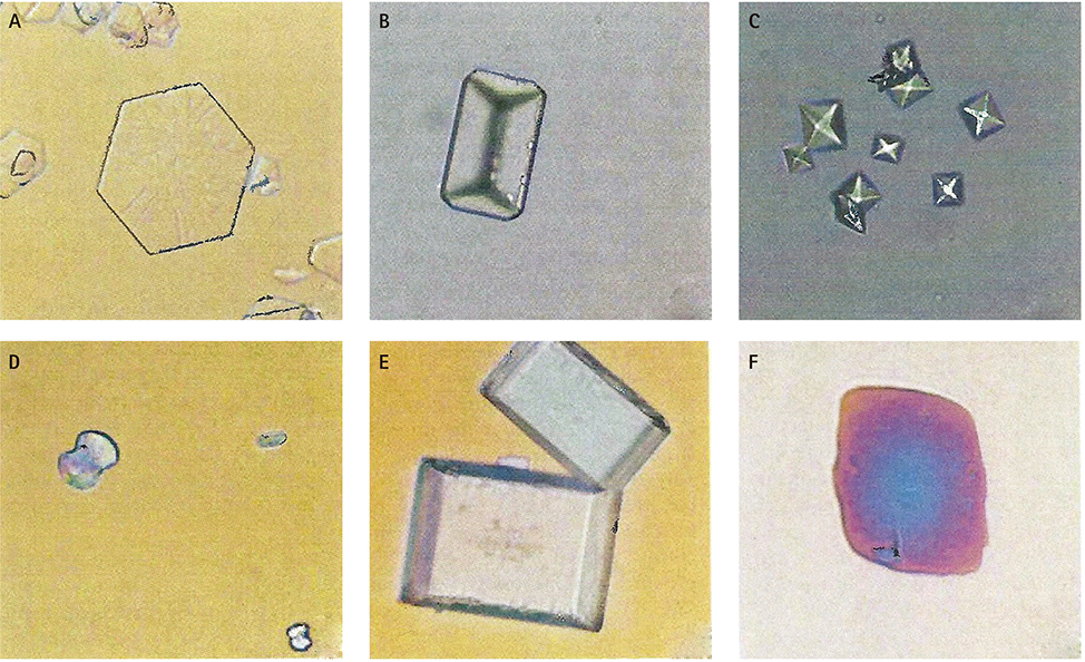
Figure 1 Type of stones. Light microscopy of urine crystals. (A) Hexagonal cystine crystal (200X); (B) coffin-lid shaped struvite crystals (200X); (C) pyramid-shaped calcium oxalate dehydrate crystals (200X); (D) dumbbell-shaped calcium oxalate monohydrate crystal (400X); (E) rectangular uric acid crystals (400X); and (F) rhomboidal uric acid crystals (400X). Reprinted with permission by Elsevier and reference [12].
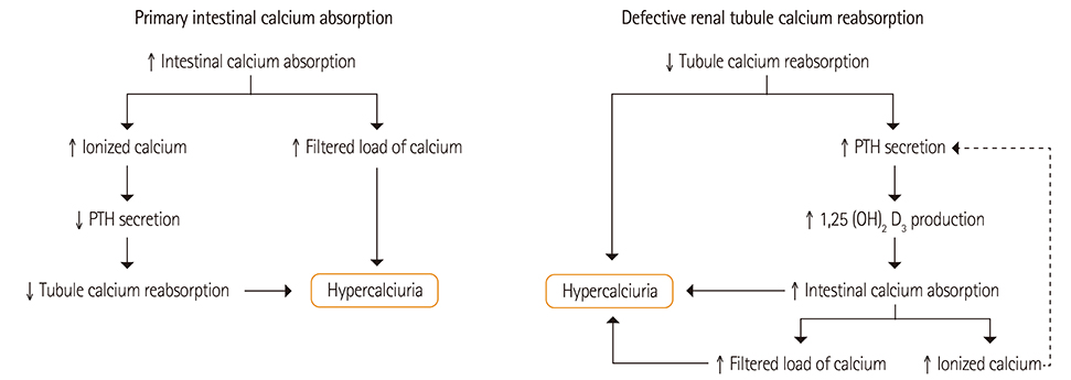
Figure 2 Model of idiopathic hypercalciuria.
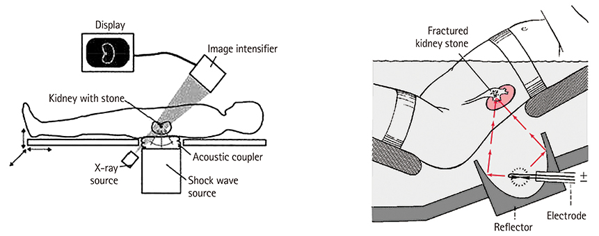
Figure 3 Extracorporeal shock wave lithotripsy (ESWL).
Cited by 1 articles
-
A prediction model of Nephrolithiasis Risk: A population-based cohort study in Korea
David Mukasa, Joohon Sung
Investig Clin Urol. 2020;61(2):188-199. doi: 10.4111/icu.2020.61.2.188.
Reference
-
1. Stamatelou KK, Francis ME, Jones CA, Nyberg LM, Curhan GC. Time trends in reported prevalence of kidney stones in the United States: 1976-1994. Kidney Int. 2003; 63:1817–1823.
Article2. Scales CD Jr, Smith AC, Hanley JM, Saigal CS. Urologic Diseases in America Project. Prevalence of kidney stones in the United States. Eur Urol. 2012; 62:160–165.
Article3. Curhan GC, Willett WC, Rimm EB, Speizer FE, Stampfer MJ. Body size and risk of kidney stones. J Am Soc Nephrol. 1998; 9:1645–1652.
Article4. Stapleton AE, Dziura J, Hooton TM, Cox ME, Yarova-Yarovaya Y, Chen S, Gupta K. Recurrent urinary tract infection and urinary Escherichia coli in women ingesting cranberry juice daily: a randomized controlled trial. Mayo Clin Proc. 2012; 87:143–150.
Article5. Goldfarb DS. In the clinic. Nephrolithiasis. Ann Intern Med. 2009; 151:ITC2.6. Pearle MS. Prevention of nephrolithiasis. Curr Opin Nephrol Hypertens. 2001; 10:203–209.
Article7. Pearle MS, Calhoun EA, Curhan GC. Urologic Diseases of America Project. Urologic diseases in America project: urolithiasis. J Urol. 2005; 173:848–857.
Article8. Asplin J, Parks J, Lingeman J, Kahnoski R, Mardis H, Lacey S, Goldfarb D, Grasso M, Coe F. Supersaturation and stone composition in a network of dispersed treatment sites. J Urol. 1998; 159:1821–1825.
Article9. Asplin JR. Nephrolithiasis: introduction. Semin Nephrol. 2008; 28:97–98.
Article10. Coe FL, Wise H, Parks JH, Asplin JR. Proportional reduction of urine supersaturation during nephrolithiasis treatment. J Urol. 2001; 166:1247–1251.
Article11. Borghi L, Guerra A, Meschi T, Briganti A, Schianchi T, Allegri F, Novarini A. Relationship between supersaturation and calcium oxalate crystallization in normals and idiopathic calcium oxalate stone formers. Kidney Int. 1999; 55:1041–1050.
Article12. Asplin JR. Evaluation of the kidney stone patient. Semin Nephrol. 2008; 28:99–110.
Article13. Finkielstein VA, Goldfarb DS. Strategies for preventing calcium oxalate stones. CMAJ. 2006; 174:1407–1409.
Article14. Kok DJ, Iestra JA, Doorenbos CJ, Papapoulos SE. The effects of dietary excesses in animal protein and in sodium on the composition and the crystallization kinetics of calcium oxalate monohydrate in urines of healthy men. J Clin Endocrinol Metab. 1990; 71:861–867.
Article15. Lemann J Jr, Worcester EM, Gray RW. Hypercalciuria and stones. Am J Kidney Dis. 1991; 17:386–391.
Article16. Worcester EM, Coe FL. Nephrolithiasis. Prim Care. 2008; 35:369–391. vii
Article17. Worcester EM, Coe FL. New insights into the pathogenesis of idiopathic hypercalciuria. Semin Nephrol. 2008; 28:120–132.
Article18. Asplin JR. Hyperoxaluric calcium nephrolithiasis. Endocrinol Metab Clin North Am. 2002; 31:927–949.
Article19. Borghi L, Meschi T, Maggiore U, Prati B. Dietary therapy in idiopathic nephrolithiasis. Nutr Rev. 2006; 64:301–312.
Article20. Borghi L, Schianchi T, Meschi T, Guerra A, Allegri F, Maggiore U, Novarini A. Comparison of two diets for the prevention of recurrent stones in idiopathic hypercalciuria. N Engl J Med. 2002; 346:77–84.
Article21. Curhan GC. Dietary calcium, dietary protein, and kidney stone formation. Miner Electrolyte Metab. 1997; 23:261–264.22. Curhan GC, Curhan SG. Dietary factors and kidney stone formation. Compr Ther. 1994; 20:485–489.23. Curhan GC, Willett WC, Rimm EB, Stampfer MJ. A prospective study of the intake of vitamins C and B6, and the risk of kidney stones in men. J Urol. 1996; 155:1847–1851.
Article24. Taylor EN, Curhan GC. Oxalate intake and the risk for nephrolithiasis. J Am Soc Nephrol. 2007; 18:2198–2204.
Article25. Curhan GC, Willett WC, Rimm EB, Stampfer MJ. A prospective study of dietary calcium and other nutrients and the risk of symptomatic kidney stones. N Engl J Med. 1993; 328:833–838.
Article26. Dawson CH, Tomson CR. Kidney stone disease: pathophysiology, investigation and medical treatment. Clin Med. 2012; 12:467–471.
Article27. Eisner BH, Goldfarb DS. A nomogram for the prediction of kidney stone recurrence. J Am Soc Nephrol. 2014; 25:2685–2687.
Article28. Asplin JR, Coe FL. Hyperoxaluria in kidney stone formers treated with modern bariatric surgery. J Urol. 2007; 177:565–569.
Article29. Lieske JC, Kumar R, Collazo-Clavell ML. Nephrolithiasis after bariatric surgery for obesity. Semin Nephrol. 2008; 28:163–173.
Article30. Cameron MA, Sakhaee K. Uric acid nephrolithiasis. Urol Clin North Am. 2007; 34:335–346.
Article31. Coe FL, Parks JH, Asplin JR. The pathogenesis and treatment of kidney stones. N Engl J Med. 1992; 327:1141–1152.
Article32. Moran ME. Uric acid stone disease. Front Biosci. 2003; 8:s1339–s1355.
Article33. Beara-Lasic L, Pillinger MH, Goldfarb DS. Advances in the management of gout: critical appraisal of febuxostat in the control of hyperuricemia. Int J Nephrol Renovasc Dis. 2010; 3:1–10.34. Kenny JE, Goldfarb DS. Update on the pathophysiology and management of uric acid renal stones. Curr Rheumatol Rep. 2010; 12:125–129.
Article35. Curhan GC, Taylor EN. 24-h uric acid excretion and the risk of kidney stones. Kidney Int. 2008; 73:489–496.
Article36. Fattah H, Hambaroush Y, Goldfarb DS. Cystine nephrolithiasis. Transl Androl Urol. 2014; 3:228–233.37. Mattoo A, Goldfarb DS. Cystinuria. Semin Nephrol. 2008; 28:181–191.
Article38. Flannigan R, Choy WH, Chew B, Lange D. Renal struvite stones--pathogenesis, microbiology, and management strategies. Nat Rev Urol. 2014; 11:333–341.
Article39. Romero-Vargas L, Barba Abad J, Rosell Costa D, Pascual Piedrola JI. Staghorn stones in renal graft. Presentation on two cases report and review the bibliography. Arch Esp Urol. 2014; 67:650–653.40. Trinchieri A. Urinary calculi and infection. Urologia. 2014; 81:93–98.
Article41. Eisner BH, McQuaid JW, Hyams E, Matlaga BR. Nephrolithiasis: what surgeons need to know. AJR Am J Roentgenol. 2011; 196:1274–1278.
Article42. Curhan GC, Willett WC, Rimm EB, Stampfer MJ. Family history and risk of kidney stones. J Am Soc Nephrol. 1997; 8:1568–1573.
Article43. Parks JH, Coe FL, Evan AP, Worcester EM. Clinical and laboratory characteristics of calcium stone-formers with and without primary hyperparathyroidism. BJU Int. 2009; 103:670–678.
Article44. Parks JH, Goldfisher E, Asplin JR, Coe FL. A single 24-hour urine collection is inadequate for the medical evaluation of nephrolithiasis. J Urol. 2002; 167:1607–1612.
Article45. Fakheri RJ, Goldfarb DS. Association of nephrolithiasis prevalence rates with ambient temperature in the United States: a re-analysis. Kidney Int. 2009; 76:798.
Article46. Fakheri RJ, Goldfarb DS. Ambient temperature as a contributor to kidney stone formation: implications of global warming. Kidney Int. 2011; 79:1178–1185.
Article47. Taylor EN, Curhan GC. Fructose consumption and the risk of kidney stones. Kidney Int. 2008; 73:207–212.
Article48. Taylor EN, Curhan GC. Dietary calcium from dairy and nondairy sources, and risk of symptomatic kidney stones. J Urol. 2013; 190:1255–1259.
Article49. Taylor EN, Stampfer MJ, Curhan GC. Dietary factors and the risk of incident kidney stones in men: new insights after 14 years of follow-up. J Am Soc Nephrol. 2004; 15:3225–3232.
Article50. Ticinesi A, Nouvenne A, Maalouf NM, Borghi L, Meschi T. Salt and nephrolithiasis. Nephrol Dial Transplant. Forthcoming 2014.
Article51. Atan L, Andreoni C, Ortiz V, Silva EK, Pitta R, Atan F, Srougi M. High kidney stone risk in men working in steel industry at hot temperatures. Urology. 2005; 65:858–861.
Article52. Borghi L, Meschi T, Amato F, Briganti A, Novarini A, Giannini A. Urinary volume, water and recurrences in idiopathic calcium nephrolithiasis: a 5-year randomized prospective study. J Urol. 1996; 155:839–843.
Article53. Goldfarb DS, Coe FL. Beverages, diet, and prevention of kidney stones. Am J Kidney Dis. 1999; 33:398–400.
Article54. Nouvenne A, Meschi T, Guerra A, Allegri F, Prati B, Borghi L. Dietary treatment of nephrolithiasis. Clin Cases Miner Bone Metab. 2008; 5:135–141.55. Taylor EN, Curhan GC. Diet and fluid prescription in stone disease. Kidney Int. 2006; 70:835–839.
Article56. Ticinesi A, Nouvenne A, Borghi L, Meschi T. Water and other fluids in nephrolithiasis: state of the art and future challenges. Crit Rev Food Sci Nutr. Forthcoming 2015.
Article57. Worcester EM, Coe FL, Evan AP, Bergsland KJ, Parks JH, Willis LR, Clark DL, Gillen DL. Evidence for increased postprandial distal nephron calcium delivery in hypercalciuric stone-forming patients. Am J Physiol Renal Physiol. 2008; 295:F1286–F1294.
Article58. Taylor EN, Curhan GC. Determinants of 24-hour urinary oxalate excretion. Clin J Am Soc Nephrol. 2008; 3:1453–1460.
Article59. Taylor EN, Stampfer MJ, Curhan GC. Diabetes mellitus and the risk of nephrolithiasis. Kidney Int. 2005; 68:1230–1235.
Article60. Tasca A. Metabolic syndrome and bariatric surgery in stone disease etiology. Curr Opin Urol. 2011; 21:129–133.
Article61. Jiang J, Knight J, Easter LH, Neiberg R, Holmes RP, Assimos DG. Impact of dietary calcium and oxalate, and Oxalobacter formigenes colonization on urinary oxalate excretion. J Urol. 2011; 186:135–139.
Article62. Lieske JC, Goldfarb DS, De Simone C, Regnier C. Use of a probiotic to decrease enteric hyperoxaluria. Kidney Int. 2005; 68:1244–1249.63. Ashby RA, Sleet RJ. The role of citrate complexes in preventing urolithiasis. Clin Chim Acta. 1992; 210:157–165.
Article64. Kessler T, Hesse A. Cross-over study of the influence of bicarbonate-rich mineral water on urinary composition in comparison with sodium potassium citrate in healthy male subjects. Br J Nutr. 2000; 84:865–871.
Article65. Kessler T, Jansen B, Hesse A. Effect of blackcurrant-, cranberry- and plum juice consumption on risk factors associated with kidney stone formation. Eur J Clin Nutr. 2002; 56:1020–1023.
Article66. Mahan LK, Escott-Stump S. Krause's food & nutrition therapy. 12th ed. St. Louis (MO): Elsevier Saunders;2008.67. Nouvenne A, Meschi T, Prati B, Guerra A, Allegri F, Vezzoli G, Soldati L, Gambaro G, Maggiore U, Borghi L. Effects of a low-salt diet on idiopathic hypercalciuria in calcium-oxalate stone formers: a 3-mo randomized controlled trial. Am J Clin Nutr. 2010; 91:565–570.
Article68. Taylor EN, Fung TT, Curhan GC. DASH-style diet associates with reduced risk for kidney stones. J Am Soc Nephrol. 2009; 20:2253–2259.
Article69. Taylor EN, Stampfer MJ, Mount DB, Curhan GC. DASH-style diet and 24-hour urine composition. Clin J Am Soc Nephrol. 2010; 5:2315–2322.
Article70. Cameron MA, Maalouf NM, Adams-Huet B, Moe OW, Sakhaee K. Urine composition in type 2 diabetes: predisposition to uric acid nephrolithiasis. J Am Soc Nephrol. 2006; 17:1422–1428.
Article71. Massey LK, Kynast-Gales SA. Diets with either beef or plant proteins reduce risk of calcium oxalate precipitation in patients with a history of calcium kidney stones. J Am Diet Assoc. 2001; 101:326–331.
Article72. Sorensen MD, Kahn AJ, Reiner AP, Tseng TY, Shikany JM, Wallace RB, Chi T, Wactawski-Wende J, Jackson RD, O'Sullivan MJ, Sadetsky N, Stoller ML. WHI Working Group. Impact of nutritional factors on incident kidney stone formation: a report from the WHI OS. J Urol. 2012; 187:1645–1649.
Article73. Noori N, Honarkar E, Goldfarb DS, Kalantar-Zadeh K, Taheri M, Shakhssalim N, Parvin M, Basiri A. Urinary lithogenic risk profile in recurrent stone formers with hyperoxaluria: a randomized controlled trial comparing DASH (Dietary Approaches to Stop Hypertension)-style and low-oxalate diets. Am J Kidney Dis. 2014; 63:456–463.
Article74. Grases F, March JG, Prieto RM, Simonet BM, Costa-Bauzá A, García-Raja A, Conte A. Urinary phytate in calcium oxalate stone formers and healthy people-dietary effects on phytate excretion. Scand J Urol Nephrol. 2000; 34:162–164.
Article75. Koff SG, Paquette EL, Cullen J, Gancarczyk KK, Tucciarone PR, Schenkman NS. Comparison between lemonade and potassium citrate and impact on urine pH and 24-hour urine parameters in patients with kidney stone formation. Urology. 2007; 69:1013–1016.
Article76. Grases F, Costa-Bauzá A. Phytate (IP6) is a powerful agent for preventing calcifications in biological fluids: usefulness in renal lithiasis treatment. Anticancer Res. 1999; 19:3717–3722.77. Grases F, Costa-Bauza A, Prieto RM. Renal lithiasis and nutrition. Nutr J. 2006; 5:23.
Article78. Grases F, Isern B, Sanchis P, Perello J, Torres JJ, Costa-Bauza A. Phytate acts as an inhibitor in formation of renal calculi. Front Biosci. 2007; 12:2580–2587.
Article79. Teichman JM. Clinical practice. Acute renal colic from ureteral calculus. N Engl J Med. 2004; 350:684–693.80. Pennington JA, Spungen JS. Bowes & Church's food values of portions commonly used. 19th ed. Philadelphia (PA): Lippincott Williams & Wilkins;2010.81. Chai W, Liebman M. Oxalate content of legumes, nuts, and grain-based flours. J Food Compost Anal. 2005; 18:723–729.
Article82. Judprasong K, Charoenkiatkul S, Sungpuag P, Vasanachitt K, Nakjamanong Y. Total and soluble oxalate contents in Thai vegetables, cereal grains and legume seeds and their changes after cooking. J Food Compost Anal. 2006; 19:340–347.
Article83. United States Department of Agriculture Agricultural Research Service. Oxalic acid content of selected vegetables. 2005. cited 2015 July 6. Available from http://www.ars.usda.gov/Services/docs.htm?docid=9444.84. Heilberg IP, Goldfarb DS. Optimum nutrition for kidney stone disease. Adv Chronic Kidney Dis. 2013; 20:165–174.
Article
- Full Text Links
-
- Actions
-
Cited
- CITED
-
- Close
- Share
-
- Similar articles
-
- Epidermoid Cyst in the Kidney with Nephrolithiasis: A Case Report
- Impact of the Human Microbiome on Nephrolithiasis
- The Effect of Dietary Calcium and Vitamin D on Renal Stone Formation
- Oral administration of oxalate decarboxylase prevents hyperoxaluria and renal calcium oxalate stone formation in ethylene glycolinduced nephrolithiasis rats
- Epidemiology and economics of nephrolithiasis
