Dual Pulsed-Wave Doppler Tracing of Right Ventricular Inflow and Outflow: Single Cardiac Cycle Right Ventricular Tei Index and Evaluation of Right Ventricular Function
- Affiliations
-
- 1Department of Medicine, Samsung Medical Center, Sungkyunkwan University School of Medicine, Seoul, Korea. parksmc@gmail.com
- 2Department of Pediatrics, Samsung Medical Center, Sungkyunkwan University School of Medicine, Seoul, Korea.
- KMID: 2145531
- DOI: http://doi.org/10.4070/kcj.2010.40.8.391
Abstract
- BACKGROUND AND OBJECTIVES
The reliability and usefulness of the right ventricular (RV) Tei index (RTX) remains controversial because it has not been possible to simultaneously measure RV inflow and outflow. However, dual pulsed-wave Doppler (DPD) enables flow velocities to be obtained at different sampling sites simultaneously. In this study we evaluated the feasibility and reliability of RTX values obtained by DPD (RTX(DPD)).
SUBJECTS AND METHODS
Forty-one patients who underwent cardiac catheterization and echocardiography for RV volume or pressure overloading conditions were evaluated. Symptom-limited exercise treadmill testing with expired gas analysis was performed and maximal exercise capacity was measured.
RESULTS
RTX by conventional flow Doppler (RTX(CFD), 0.262+/-0.164) was similar to RTX(DPD) (0.253+/-0.117, p=NS), whereas RTX by tissue Doppler echocardiography (RTX(TDE), 0.447+/-0.125) was significantly larger than RTX(DPD) (p<0.001). Based on multiple regression analysis, maximal exercise capacity was independently related to RTX(DPD) (beta=-0.60, p<0.001), mid-RV dimension (beta=-0.26, p=0.012), left ventricular ejection fraction (beta=0.22, p=0.023), and early diastolic tricuspid annular velocity (beta=0.21, p=0.048).
CONCLUSION
It is feasible and reliable to evaluate RV function using RTX(DPD) values. However, to evaluate the clinical usefulness of RTX(DPD), additional studies are required with a large number of patients and long-term follow-up.
MeSH Terms
Figure
-
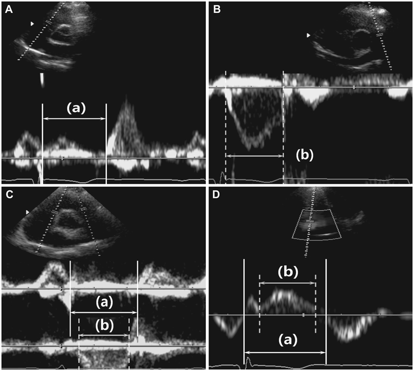
Fig. 1 Measurement of right ventricular Tei index (RTX) by (A and B) the conventional flow Doppler method (RTXCFD), (C) the dual pulsed-wave Doppler method (RTXDPD), and by (D) tissue Doppler echocardiography (RTXTDE). RTX was defined as [(a)-(b)]/(b), where (a) is the time from tricuspid valve inflow cessation to onset for RTXCFD and RTXDPD and time from the end of A'TV to the onset of E'TV for RTXTDE, and (b) is the pulmonary ejection time for RTXCFD and RTXDPD or the duration of S'TV for RTXTDE. A'TV: late diastolic tricuspid annular velocity, E'TV: early diastolic tricuspid annular velocity, S'TV: systolic tricuspid annular velocity.

Fig. 2 Correlation (left column) and Altman-Bland plots (right column) between right ventricular Tei indexes (RTX) using conventional flow Doppler (CFD) and dual pulsed-wave Doppler (DPD; upper row); RTX using CFD and tissue Doppler (TDE; mid-row); and RTX using the DPD and TDE methods.
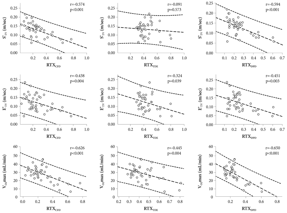
Fig. 3 Correlation plots of the right ventricular Tei index (RTX) vs. the S'TV (upper row), E'TV (mid-row), and maximal exercise capacity (lower row). Horizontal axes in the leftmost column represent RTX values determined using conventional flow Doppler (RTXCFD); the middle column represents RTX values determined using tissue Doppler echocardiography (RTXTDE); and the right column RTX values determined using the dual pulsed-wave Doppler method (RTXDPD). S'TV: systolic tricuspid annular velocity, E'TV: early diastolic tricuspid annular velocity.
Reference
-
1. Tei C, Ling LH, Hodge DO, et al. New index of combined systolic and diastolic myocardial performance: a simple and reproducible measure of cardiac function--a study in normals and dilated cardiomyopathy. J Cardiol. 1995. 26:357–366.2. Dujardin KS, Tei C, Yeo TC, Hodge DO, Rossi A, Seward JB. Prognostic value of a Doppler index combining systolic and diastolic performance in idiopathic-dilated cardiomyopathy. Am J Cardiol. 1998. 82:1071–1076.3. Tei C, Dujardin KS, Hodge DO, Kyle RA, Tajik AJ, Seward JB. Doppler index combining systolic and diastolic myocardial performance: clinical value in cardiac amyloidosis. J Am Coll Cardiol. 1996. 28:658–664.4. Sohn IS, Kang HS, Kim SJ, et al. Tissue Doppler image-derived myocardial performance (Tei index) as a simple assessment of global cardiac function in adults. Korean Circ J. 2005. 35:315–321.5. Cho K, Park J, Lee C, et al. Isolated and combined influences of diabetes and hypertension on the myocardial function and geometry. Korean Circ J. 2006. 36:411–417.6. Voon WC, Su HM, Yen HW, Lin TH, Lai WT, Sheu SH. Left ventricular Tei index: comparison between flow and tissue Doppler analyses. Echocardiography. 2005. 22:730–735.7. Harada K, Tamura M, Toyono M, Yasuoka K. Comparison of the right ventricular Tei index by tissue Doppler imaging to that obtained by pulsed Doppler in children without heart disease. Am J Cardiol. 2002. 90:566–569.8. Grignola JC, Gines F, Guzzo D. Comparison of the Tei index with invasive measurements of right ventricular function. Int J Cardiol. 2006. 113:25–33.9. Roberson DA, Cui W. Right ventricular Tei index in children: effect of method, age, body surface area, and heart rate. J Am Soc Echocardiogr. 2007. 20:764–770.10. Schiller NB, Shah PM, Crawford M, et al. Recommendations for quantitation of the left ventricle by two-dimensional echocardiography. American Society of Echocardiography Committee on Standards, Subcommittee on Quantitation of Two-Dimensional Echocardiograms. J Am Soc Echocardiogr. 1989. 2:358–367.11. Pritchett AM, Jacobsen SJ, Mahoney DW, Rodeheffer RJ, Bailey KR, Redfield MM. Left atrial volume as an index of left atrial size: a population-based study. J Am Coll Cardiol. 2003. 41:1036–1043.12. Gilman G, Nelson TA, Hansen WH, Khandheria BK, Ommen SR. Diastolic function: a sonographer's approach to the essential echocardiographic measurements of left ventricular diastolic function. J Am Soc Echocardiogr. 2007. 20:199–209.
- Full Text Links
-
- Actions
-
Cited
- CITED
-
- Close
- Share
-
- Similar articles
-
- Tissue Doppler Image-Derived Myocardial Performance(Tei Index) as a Simple Assessment of Global Cardiac Function in Adults
- Assessment of Left Ventricular Diastolic Pressures with Pulmonary Venous Flow and Transmitral Inflow by Doppler Echocardiography
- Modified Tei index in patients with Kawasaki disease by tissue doppler imaging
- A Case of Ventricular Septal Defect After Acute Myocardial Infarction
- A Case of Left Ventricular Outflow Obstruction Caused by Mitral Valve Replacement
