Acinar Cell Carcinoma of the Pancreas: A Report of Two Cases with Long-term Follow-up and a Review of the Literature
- Affiliations
-
- 1Department of Surgery, Hanyang University College of Medicine, Seoul, Korea. hkpark@hanyang.ac.kr
- 2Department of Pathology, Hanyang University College of Medicine, Seoul, Korea.
- KMID: 2145035
- DOI: http://doi.org/10.4174/jkss.2010.79.4.310
Abstract
- Acinar cell carcinoma (ACC) of the pancreas is a rare malignancy making up approximately 1% of pancreatic non-endocrine malignant tumors. The common finding on computed tomography is a solitary, well-defined, heterogenous hypodense mass with enhancing capsule. ACC is a highly cellular tumor with minimal stroma and a lack of stromal desmoplasia. The accurate diagnosis of ACC cannot typically be done by histology alone but rather requires immunohistochemical staining or electron microscopy for the identification of pancreatic enzymes and zymogen granules. ACC has been considered a cancer with a poor prognosis due to frequent metastasis, a high recurrence rate, and low respectability. Surgical resection is the treatment of choice that can lead to long-term survival. ACC has a better prognosis than ductal carcinoma of the pancreas, but a worse prognosis compared to islet cell carcinoma. We report two cases of ACC with 5-year survival after surgical resection.
Keyword
MeSH Terms
Figure
-
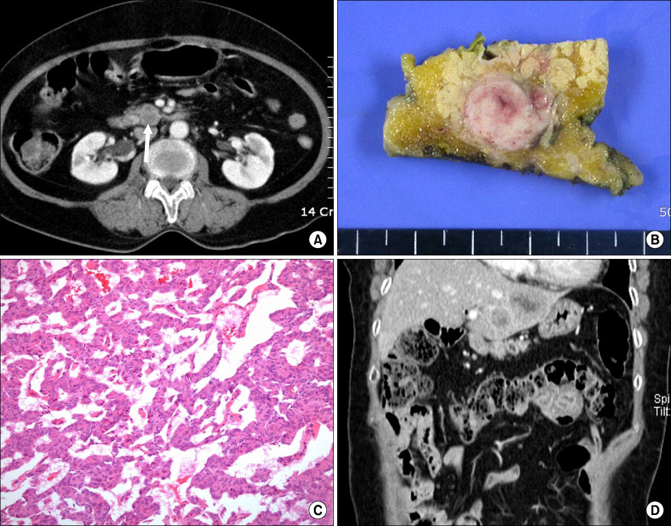
Fig. 1 (A) Contrast-enhanced axial CT scan reveals heterogenous 2 cm low-attenuated lesion (arrow) around head of pancreas (portal phase). (B) Cut surface of tumor shows relatively well-defined and grayish-white round mass. (C) Tumor demonstrates trabecular growth pattern of acinic tumor cells (H&E stain, ×200). (D) Contrast-enhanced coronal CT scans show two metastatic tumors within segment II of liver on 58 months after surgery (portal phase).
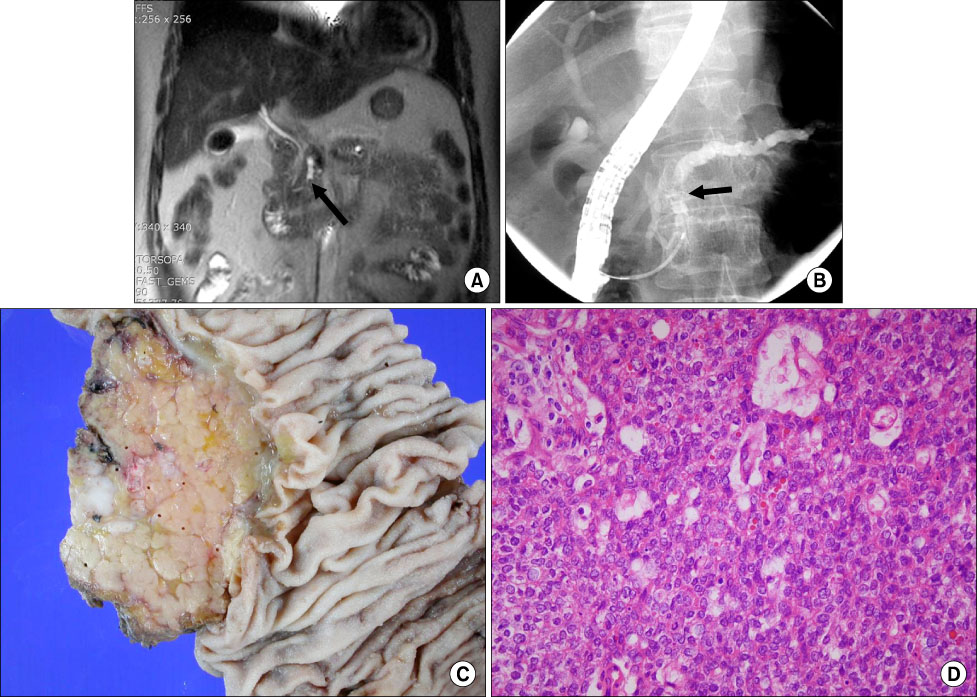
Fig. 2 (A) Magnetic resonance cholangiopancreatography shows focal stenosis (arrow) of pancreatic duct in head and diffuse duct dilatation of body and tail. (B) Endoscopic retrograde cholangiopancreatography findings shows filling defect (arrow) of pancreatic duct between head and body, as well as diffuse dilatation of body and tail of pancreatic duct. (C) Pancreatic head reveals an ill-defined whitish multi-nodular solid mass, measuring 1.2 cm in diameter. (D) Microscopic view demonstrates mostly solid and focally tubular tumor nests. Tumor cells have relatively uniform oval nuclei and moderate amount eosinophilic granular cytoplasm (H&E stain, ×400).
Reference
-
1. Klimstra DS, Heffess CS, Oertel JE, Rosai J. Acinar cell carcinoma of the pancreas. A clinicopathologic study of 28 cases. Am J Surg Pathol. 1992. 16:815–837.2. Caruso RA, Inferrera A, Tuccari G, Barresi G. Acinar cell carcinoma of the pancreas. A histologic, immunocytochemical and ultrastructural study. Histol Histopathol. 1994. 9:53–58.3. Seth AK, Argani P, Campbell KA, Cameron JL, Pawlik TM, Schulick RD, et al. Acinar cell carcinoma of the pancreas: an institutional series of resected patients and review of the current literature. J Gastrointest Surg. 2008. 12:1061–1067.4. Kitagami H, Kondo S, Hirano S, Kawakami H, Egawa S, Tanaka M. Acinar cell carcinoma of the pancreas: clinical analysis of 115 patients from Pancreatic Cancer Registry of Japan Pancreas Society. Pancreas. 2007. 35:42–46.5. Schmidt CM, Matos JM, Bentrem DJ, Talamonti MS, Lillemoe KD, Bilimoria KY. Acinar cell carcinoma of the pancreas in the United States: prognostic factors and comparison to ductal adenocarcinoma. J Gastrointest Surg. 2008. 12:2078–2086.6. Ordóñez NG. Pancreatic acinar cell carcinoma. Adv Anat Pathol. 2001. 8:144–159.7. Holen KD, Klimstra DS, Hummer A, Gonen M, Conlon K, Brennan M, et al. Clinical characteristics and outcomes from an institutional series of acinar cell carcinoma of the pancreas and related tumors. J Clin Oncol. 2002. 20:4673–4678.8. Chiou YY, Chiang JH, Hwang JI, Yen CH, Tsay SH, Chang CY. Acinar cell carcinoma of the pancreas: clinical and computed tomography manifestations. J Comput Assist Tomogr. 2004. 28:180–186.9. Webb JN. Acinar cell neoplasms of the exocrine pancreas. J Clin Pathol. 1977. 30:103–112.10. Oh JY, Nam KJ, Choi JC, Suh SB, Lee KN, Park BH, et al. CT findings of acinar cell carcinoma of the pancreas: focused on spiral CT findings. J Korean Radiol Soc. 2001. 45:29–34.
- Full Text Links
-
- Actions
-
Cited
- CITED
-
- Close
- Share
-
- Similar articles
-
- A Case of Acinar Cell Carcinoma of the Pancreas
- Acinar Cell Carcinoma of the Pancreas: A case report
- Clinical Study of Acinar Cell Carcinomas of the Pancreas: Our 5 cases and a review of 5 cases reported in Korea
- Mixed acinar-endocrine carcinoma of the pancreas: a case report
- Acinar Cell Carcinoma of the Pancreas: A Case Report
