J Korean Surg Soc.
2013 Feb;84(2):114-117. 10.4174/jkss.2013.84.2.114.
Primary mesenteric carcinoid tumor
- Affiliations
-
- 1Department of Surgery, St. Vincent's Hospital, The Catholic University of Korea College of Medicine, Suwon, Korea. hj@catholic.ac.kr
- 2Department of Hospital Pathology, St. Vincent's Hospital, The Catholic University of Korea College of Medicine, Suwon, Korea.
- 3Department of Radiology, St. Vincent's Hospital, The Catholic University of Korea College of Medicine, Suwon, Korea.
- KMID: 2144999
- DOI: http://doi.org/10.4174/jkss.2013.84.2.114
Abstract
- Primary mesenteric carcinoid tumor is very rare, although secondary mesenteric involvement is common, reported as 40% to 80%. And distant metastasis rate reported as 80% to 90%, when the size is larger than 2 cm. We present a case of very rare primary mesenteric carcinoid tumor showing benign character though large size. The patient visited St. Vincent's Hospital, The Catholic University of Korea with increasing palpable abdominal mass. At laparotomy, a well encapsulated mass arising from the mesentery near the ligament of Treitz was found without any adjacent organ invasion or distant metastasis. The mass was measured as 8.2 x 7.3 cm and histopathologically benign character. At 11 months of follow up, the patient was recurrence free.
Keyword
MeSH Terms
Figure
-
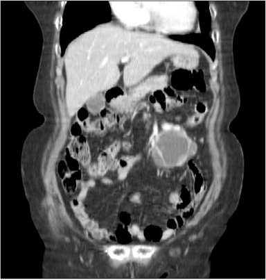
Fig. 1 Computed tomography scan shows well-defined and complex density mass in mesentery or adjacent proximal jejunum.
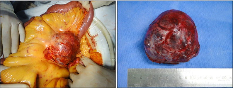
Fig. 2 Well-capsulated mass originated near the ligament of Treitz.
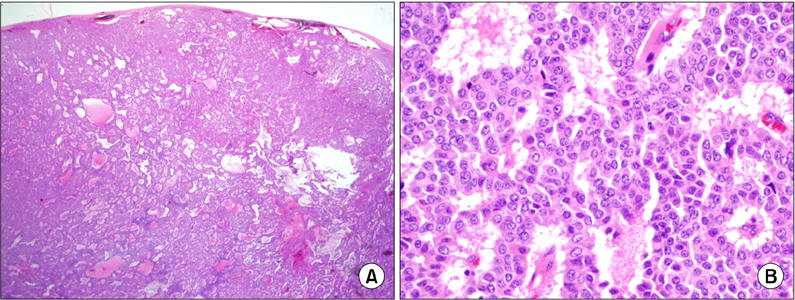
Fig. 3 (A) Cells composing tumor are arranged in trabecular pattern and well encapsulated (H&E, ×40). (B) Cellular nucleus has coarsely granular chromatin pattern without mitosis (H&E, ×400).
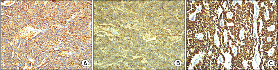
Fig. 4 Tumor expresses strong positivity in immunohisochemical stain with synaptophysin (A), CD56 (B), and cytokeratin (C) (×200).
Reference
-
1. Yamanuha J, Ballinger R, Coon D, Navin J. Carcinoid tumor presenting as a primary mesenteric mass: a case report and review of the literature. Hawaii Med J. 2009. 68:137–139.2. Karahan OI, Kahriman G, Yikilmaz A, Ozkan M, Bayram F. Gastrointestinal carcinoid tumors in rare locations: imaging findings. Clin Imaging. 2006. 30:278–282.3. Woodside KJ, Townsend CM Jr, Mark Evers B. Current management of gastrointestinal carcinoid tumors. J Gastrointest Surg. 2004. 8:742–756.4. Akerstrom G, Hellman P, Hessman O, Osmak L. Management of midgut carcinoids. J Surg Oncol. 2005. 89:161–169.5. de Vries H, Verschueren RC, Willemse PH, Kema IP, de Vries EG. Diagnostic, surgical and medical aspect of the midgut carcinoids. Cancer Treat Rev. 2002. 28:11–25.
- Full Text Links
-
- Actions
-
Cited
- CITED
-
- Close
- Share
-
- Similar articles
-
- Primary Carcinoid Tumor Arising in a Mature Teratoma of the Testis: A Case Report
- A Case of Primary Carcinoid Tumor of the Urinary Bladder
- Primary Carcinoid Tumor of the Testis: A case report
- Primary Renal Carcinoid Tumor
- MRI Features with Pathologic Correlation of Primary Ovarian Carcinoid: A Case Report
