Korean J Urol.
2006 Oct;47(10):1130-1132. 10.4111/kju.2006.47.10.1130.
Fibrous Pseudotumor in the Testicular Tunica
- Affiliations
-
- 1Department of Urology, College of Midicine, Dong-a University College of Medicine, Busan, Korea. urobone@korea.com
- 2Seomyon Medical Center, Busan, Korea.
- 3Department of Pathology, College of Midicine, Dong-a University College of Medicine, Busan, Korea.
- KMID: 2139819
- DOI: http://doi.org/10.4111/kju.2006.47.10.1130
Abstract
- Fibrous pseudotumor of the testicular tunics is uncommon lesion. They typically arise as painless scrotal masses that may be associated with a hydrocele or history of trauma or infection. Two-thirds involve the tunica vaginalis testis, with infrequent involvement of other scrotal structures. Once excised, these lesions behave in a benign fashion. Typically, these masses are multinodular, but in rare cases they are diffuse, band-like myofibroblastic proliferations that encase the testis. We report here on a case of fibrous pseudotumor of the tunica vaginalis in 76 year-old patient. (Korean J Urol 2006;47:1130-1132)
Keyword
MeSH Terms
Figure
-

Fig. 1 Scrotal ultrasonography findings. (A) A slightly hyperechoic lesion that totally replaces the left testis. (B) Doppler ultrasonography shows an increase of vascularity.
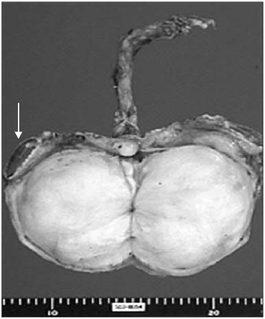
Fig. 2 Gross findings. A large nodular paratesticular mass enclosed in tunica albuginea is compressing the atrophied testis (arrow). The mass measures 10×7cm in dimension and it shows whitish homogenous fibrotic cut surface with slightly firm consistency.

Fig. 3 Microscopic findings. (A) The mass is composed of dense fibrocollageneous areas. (B) The mass is composed of loose, myxoid areas. A few atypical fibroblasts with enlarged and hyperchromatic nuclei are noted, but without mitosis (H&E stain, ×100).
Reference
-
1. Ulbright TM, Amin MB, Young RH. Ulbright TM, editor. Tumors of testis, adnexa, spermatic cord, and scrotum. Atlas of tumor pathology. 1999. 3rd ed. Washington DC: Armed Forces Institutes of pathology;317–319.2. Mostifi FK, Price EB. Tumors of the Male Genital System. 1973. Washington DC: Armed Forces Institute of Pathology;151–154.3. Yang LC, Kwun MH, Kim HH, Kim KS, Noh JH, Kim SI, et al. Fibrous pseudotumor of paratesticular region. Korean J Urol. 2003. 44:941–943.4. Paik SS, Kim NH, Ko YH, Park MH. Fibrous pseudotumor of the testicular tunics. Korean J Pathol. 1995. 29:533–535.5. Kim NR, Ha SY, Chung JG, Han JH. Inflammatory pseudotumor of the paratesticular area. Korean J Pathol. 2004. 38:208–211.6. Tobias-machado M, Correa Lopes Neto A, Heloisa Simardi L, Borrelli M, Wroclawski ER. Fibrous pseudotumor of tunica vaginalis and epididymis. Urology. 2000. 56:670–672.7. Williams G, Banerjee R. Paratesticular tumors. Br J Urol. 1969. 41:332–339.8. Jones MA, Young RH, Scully RE. Benign fibromatous tumors of the testis and paratesticular region: a report of 9 cases with a proposed classification of fibromatous tumors and tumor-like lesions. Am J Surg Pathol. 1997. 21:296–305.9. Sonmez K, Turkyilmaz Z, Boyacioglu M, Edali MN, Ozen O, Basaklar AC, et al. Diffuse fibrous proliferation of tunica vaginalis associated with testicular infarction: a case report. J Pediatr Surg. 2001. 36:1057–1058.10. Lai FM, Allen PW, Chan LW, Chan PS, Cooper JE, Mackenzie TM. Aggressive fibromatosis of the spermatic cord. A typical lesion in a "new" location. Am J Clin Pathol. 1995. 104:403–407.
- Full Text Links
-
- Actions
-
Cited
- CITED
-
- Close
- Share
-
- Similar articles
-
- Fibrous Pseudotumor of the Testicular Tunics: Two case reports
- A Case of Fibrous Pseudotumor of Testicular Tunic
- Fibrous Pseudotumor of Testicular Tunic: A Case Report
- Ultrasonographic Findings of Fibrous Pseudotumor Arising from the Tunica Vaginalis Testis: A Case Report
- Ultrasonographic and Magnetic Resonance Imaging Findings of Fibrous Pseudotumor of the Tunica Vaginalis: A Case Report
