Ultrasonographic and Magnetic Resonance Imaging Findings of Fibrous Pseudotumor of the Tunica Vaginalis: A Case Report
- Affiliations
-
- 1Department of Radiology, College of Medicine, Yeungnam University, Daegu, Korea. jhcho@yu.ac.kr
- 2Department of Pathology, College of Medicine, Yeungnam University, Daegu, Korea.
- KMID: 1819783
- DOI: http://doi.org/10.3348/jksr.2014.71.2.75
Abstract
- A fibrous pseudotumor is a rare benign fibroproliferative disease of the paratesticular tissues, usually originating from the tunica vaginalis. A 33-year-old man with a painless palpable mass on the right scrotum visited our institution. On ultrasonography (US), multiple well-defined isoechoic nodular lesions with a small amount of vascularity were seen along the tunica vaginalis. Some of the nodules showed posterior shadowing. On magnetic resonance imaging (MRI), the lesions showed dark signal intensity on the T2-weighted image and slightly low signal intensity on the T1-weighted image. The dynamic contrast-enhanced MRI showed a mild and progressive enhancement which was persistent on the delayed phase. After surgical excision, the mass was pathologically confirmed as fibrous pseudotumor. Herein we report a case of fibrous pseudotumor of the tunica vaginalis, which was diagnosed with both US and MRI findings.
Figure
-
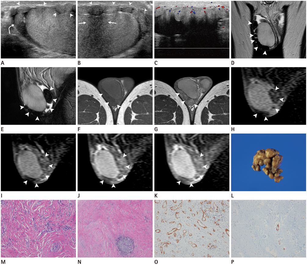
Fig. 1 A 33-year-old man with fibrous pseudotumor of the tunica vaginalis in right scrotum. A, B. Longitudinal scan of gray-scale ultrasonography (US) at the level of upper (A) and lower poles (B) of the testis shows multiple iso- to slightly hypoechoic nodular lesions (arrowheads) attached at the tunica vaginalis on the anterior aspect of the testis and minimal amount of fluid collection in the tunical sac. One of the nodules shows posterior shadowing without calcification (arrows). Epididymal head is normally well seen (curved arrow). C. On color Doppler US, small amount of vascularity is noted in the lesion. D-G. Multiple nodular lesions (arrowheads) at the tunica vaginalis show homogeneous dark signal intensity (SI) on coronal and sagittal T2-weighted image (D, E), slightly low SI on axial T1-weighted image (T1WI) (F), and mild enhancement on contrast-enhanced axial T1WI (G). H-K. On dynamic contrast-enhanced study with sagittal fat suppressed T1WI, the lesion shows mild and progressive enhancement which persists through the delayed phase (arrowheads). Pre-contrast (H), arterial (after 30 seconds, I), venous (after 90 seconds, J), and delayed phase (after 300 seconds, K) images are presented. L. Pathologically, the gross specimen of the lesion consists of right scrotal tunica vaginalis with multiple dark brownish firm nodules on the surface. M. Microscopically, the lesion shows abundant sclerotic and fibrous tissue surrounding vascular structures (hematoxylin and eosin stain, × 100). N. The lesion also shows largely hypocellular tissue with a cluster of inflammatory cells (hematoxylin and eosin stain, × 40). O, P. On immunohistochemical stain for actin (O) and desmin (P), the tumor cells reveal negative reaction (immunohistochemistry, × 100).
Reference
-
1. Bruijnes E, Laddé BE, Dabhoiwala NF, Stukart RA. Fibrous pseudotumor of the tunica vaginalis testis. Urol Int. 1984; 39:314–317.2. Grebenc ML, Gorman JD, Sumida FK. Fibrous pseudotumor of the tunica vaginalis testis: imaging appearance. Abdom Imaging. 1995; 20:379–380.3. Tobias-machado M, Corrêa Lopes Neto A, Heloisa Simardi L, Borrelli M, Wroclawski ER. Fibrous pseudotumor of tunica vaginalis and epididymis. Urology. 2000; 56:670–672.4. Parker PM, Pugliese JM, Allen RC Jr. Benign fibrous pseudotumor of tunica vaginalis testis. Urology. 2006; 68:427.e17–427.e19.5. Krainik A, Sarrazin JL, Camparo P, Vincendeau S, Houlgatte A, Cordoliani YS. Fibrous pseudotumor of the epididymis: imaging and pathologic correlation. Eur Radiol. 2000; 10:1636–1638.6. al-Otaibi L, Whitman GJ, Chew FS. Fibrous pseudotumor of the epididymis. AJR Am J Roentgenol. 1997; 168:1586.7. Cassidy FH, Ishioka KM, McMahon CJ, Chu P, Sakamoto K, Lee KS, et al. MR imaging of scrotal tumors and pseudotumors. Radiographics. 2010; 30:665–683.8. Patel MD, Silva AC. MRI of an adenomatoid tumor of the tunica albuginea. AJR Am J Roentgenol. 2004; 182:415–417.9. Hertzberg BS, Kliewer MA, Hertzberg MA, Distell BM. Epididymal leiomyoma: sonographic features. J Ultrasound Med. 1996; 15:797–799.10. Fields JM, Russell SA, Andrew SM. Case report: ultrasound appearances of a malignant mesothelioma of the tunica vaginalis testis. Clin Radiol. 1992; 46:128–130.
- Full Text Links
-
- Actions
-
Cited
- CITED
-
- Close
- Share
-
- Similar articles
-
- Ultrasonographic Findings of Fibrous Pseudotumor Arising from the Tunica Vaginalis Testis: A Case Report
- Fibrous Pseudotumor in the Testicular Tunica
- Fibrous Pseudotumor of Paratesticular Region: A case report
- A case of primary malignant mesothelioma of tunica vaginalis testis
- A Case of Fibroma of Tunica Vaginalis
