Korean J Urol.
2006 Oct;47(10):1127-1129. 10.4111/kju.2006.47.10.1127.
Adenomatoid Tumor of the Testis with Infiltration to the Seminiferous Tubules
- Affiliations
-
- 1Department of Urology, Inje University College of Medicine, Busan, Korea. prosdoc@hanmail.net
- 2Department of Pathology, Inje University College of Medicine, Busan, Korea.
- KMID: 2139818
- DOI: http://doi.org/10.4111/kju.2006.47.10.1127
Abstract
- A 41-year-old man presented a bean sized, solid, painless left scrotal mass he'd had for 10 years. The mass was well demarcated and it showed homogeneous echogenecity on ultrasonography. Simple mass excision was performed and the specimen revealed a relatively well circumscribed mass lesion composed of dilated tubules with flattened lining cells. A focal infiltration to the seminiferous tubule and involvement of the biopsy margins were observed. The tubules had a positive reaction to the calretinin and anti-mesothelial antibody on immunohistochemical stain. Therefore, the tumor was diagnosed as adenomatoid tumor of the testis. Adenomatoid tumors of testis are rare benign neoplasms that are thought to originate from mesothelum. Most cases were reported in the epididymis, spermatic cord and testicular tunica, and rare cases were from the ejaculatory duct, prostate and adrenal gland. The findings of adenomatoid tumor infiltrating through the testis parenchyme and seminiferous tubule, like for our case, have not been previously reported in Korea.
Keyword
MeSH Terms
Figure
-
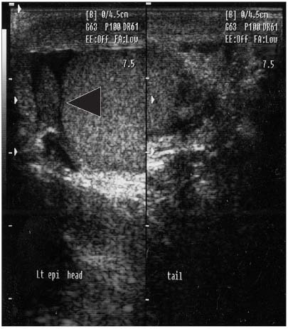
Fig. 1 Scrotal sonography. A 7×8mm sized, oval shape, well demarcated, homogeneous echogenic mass between the left epididymal head and testis (arrow head).
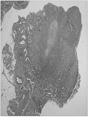
Fig. 2 The tumor lesion is located between the head of the epididymis and the rete testis with extensive fibrotic stroma (H&E, ×12.5).
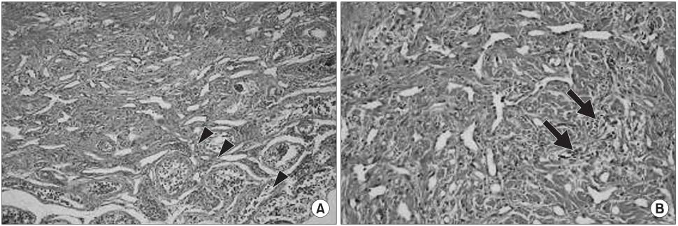
Fig. 3 The tumor is composed of dilated or stag-horn shaped tubules lined by flattened cells (narrow arrow heads), (A: H&E, ×100) and some tubules are infiltrating between the seminiferous tubules (arrow) (B: H&E, ×200).
Cited by 1 articles
-
Adenomatoid Tumor of the Spermatic Cord
Jun Shik Shin, Han Ki Yun, Yoon Hyung Lee, Eun Kyung Kwak, Sung Ryong Cho
Korean J Urol. 2008;49(7):650-652. doi: 10.4111/kju.2008.49.7.650.
Reference
-
1. Park SM, Jun IS, Hwang CH, Heo C, Lee TH, Hong SJ, et al. Adenomatoid tumor of the tunica vaginalis testis. Korean J Urol. 2004. 45:1171–1173.2. Shin MK, Kim DS, Yoon DK, Koh SK. Adenomatoid tumor of epididymis. Korean J Urol. 1986. 27:500–502.3. Oyama H, Ogawa M, Mikuriya H, Kido A, Hayashi H. Adenomatoid tumor of testicular tunica albuginea. Hinyokika Kiyo. 2001. 47:661–663.4. Delahunt B, Eble JN, King D, Bethwaite PB, Nacey JN, Thornton A. Immunohistochemical evidence for mesothelial origin of paraterticular adenomatoid tumor. Histopathology. 2000. 36:109–115.5. Han CH, Sun IC, Kwak KM, Chung WK, Ha IS, Shin OR, et al. Adenomatoid tumor of the epididymis. Korean J Urol. 2002. 43:256–258.6. Mostofi FK, Price EB. Firminger HI, editor. Tumors of the male genital system. Atalas of tumor pathology. 1973. Washington DC: Armed Forces Institute of Pathol;144–151.7. Amin MB. Selected other problematic testicular and paratesticular lesions: rete testis neoplasms and pseudotumors, mesothelial lesions and secondary tumors. Mod Pathol. 2005. 18:Suppl 2. S131–S145.8. Mucienters F, Govindrajan S, Burotto S. Immunoperoxidase study on adenomatoid tumor of the epididymis using anti-mesothelial cell serum. Cancer. 1985. 55:363–365.
