Focal Hand Dystonia Secondary to Basal Ganglia Germinoma
- Affiliations
-
- 1Department of Neurology, The Catholic University of Korea, Seoul, Korea. ks1007@catholic.ac.kr
- 2Department of Radiology, The Catholic University of Korea, Seoul, Korea.
- KMID: 2135494
- DOI: http://doi.org/10.3988/jcn.2007.3.3.150
Abstract
- Descriptions of symptomatic focal dystonia caused by focal lesions of the central nervous system (CNS) are rare in the literature. We report a 9-year-old child who experienced sudden-onset left-hand dystonia for 6 months. Brain magnetic resonance imaging showed a mass lesion involving the putamen, globus pallidus, head of caudate, and the anterior limb of the internal capsule. Histopathological and immunocytochemical examinations of the mass revealed features characteristic of malignant germinoma. CNS germinoma in the basal ganglia is very rare. Combining previous reports in the literature with the anatomical and clinical presentation of our case suggests that this phenomenon results from disruption of the pathways within and adjacent to the basal ganglia.
Keyword
MeSH Terms
Figure
-
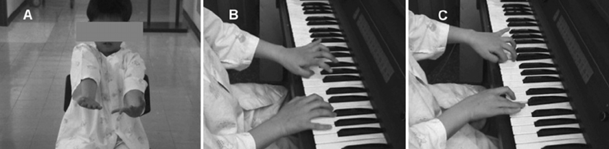
Figure 1 Photograph showing dystonia of the left hand during arm extension (A) and playing the piano (B, C).
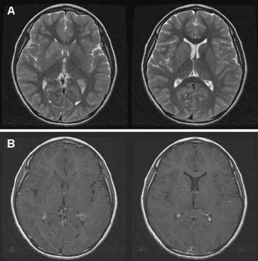
Figure 2 Magnetic resonance imaging showing an ill-defined mass lesion involving the right basal ganglia. The lesion was located in the putamen, globus pallidus, head of caudate, and the anterior limb of the internal capsule. (A) Axial T2- weighted images, and (B) axial T1-weighted contrast enhancement images.
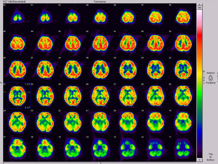
Figure 3 Positron-emission tomography using 18F-deoxyglucose demonstrated diffuse hypometabolism in the right basal ganglia, right thalamus, and right insular and temporal cortices.
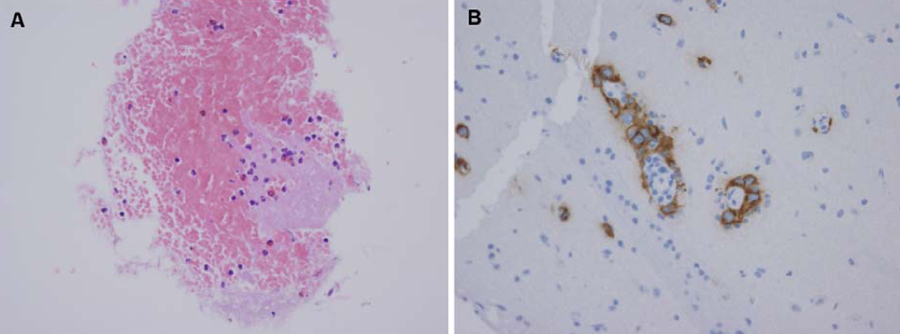
Figure 4 Histopathological and immunohistochemical findings revealed (A) large tumorous cells (germ cells) with a clear nucleus and a light-colored and vacuolated cytoplasm (hematoxylin-eosin stain, ×400) and (B) a placental alkaline phosphatase (PLAP) profile with large tumorous cells labeled for PLAP in the cell membrane and cytoplasm (×400).
Reference
-
1. Kostic VS, Stojanovic-Svetel M, Kacar A. Symptomatic dystonias associated with structural brain lesions: report of 16 cases. Can J Neurol Sci. 1996. 23:53–56.
Article2. Matsutani M, Sano K, Takakura K, Fujimaki T, Nakamura O, Funata N, et al. Primary intracranial germ cell tumors: a clinical analysis of 153 histologically verified cases. J Neurosurg. 1997. 86:446–455.
Article3. Evanson EJ, Lewis PD, Colquhoun IR. Primary germinoma of the posterior cranial fossa: a case report. Neuroradiology. 1997. 39:716–718.
Article4. Garcia-Santos JM, Torres del Rio S, Sanchez A, Martinez-Lage JF. Basal ganglia and thalamic tumours: an imaging approximation. Childs Nerv Syst. 2002. 18:412–425.
Article5. Kobayashi T, Yoshida J, Kida Y. Bilateral germ cell tumors involving the basal ganglia and thalamus. Neurosurgery. 1989. 24:579–583.
Article6. Matsumoto K, Tabuchi A, Tamesa N, Nakashima H, Ohmoto T. Primary intracranial germinoma involving the midbrain. Clin Neurol Neurosurg. 1998. 100:292–295.
Article7. Nakajima H, Iwai Y, Yamanaka K, Yasui T, Kishi H. Primary intracranial germinoma in the medulla oblongata. Surg Neurol. 2000. 53:448–451.
Article8. Yoshida K, Nakao Y, Yamamoto T, Mori K, Maeda M. Germinoma in the fourth ventricle. Acta Neurochir (Wien). 2003. 145:789–792.
Article9. Marsden CD, Obeso JA, Zarranz JJ, Lang AE. The anatomical basis of symptomatic hemidystonia. Brain. 1995. 108:463–483.
Article10. Obeso JA, Gimenez-Roldan S. Fahn S, editor. Clinicopathological correlation in symptomatic dystonia. Dystonia 2, Advances in Neurology. 1988. New York: Raven Press;113–122.11. Narbona J, Obeso JA, Tunon T, Martinez-Lage JM, Marsden CD. Hemi-dystonia secondary to localised basal ganglia tumour. J Neurol Neurosurg Psychiatry. 1984. 47:704–709.
Article12. Pettigrew LC, Jankovic J. Hemidystonia: A report of 22 patients and a review of the literature. J Neurol Neurosurg Psychiatry. 1985. 48:650–657.
Article13. Krystkowiak P, Martinat P, Defebvre L, Pruvo JP, Leys D, Destee A. Dystonia after striatopallidal and thalamic stroke: clinicoradiological correlations and pathophysiological mechanisms. J Neurol Neurosurg Psychiatry. 1998. 65:703–708.
Article14. Maillard L, Ishii K, Bushara K, Waldvogel D, Schulman AE, Hallett M. Mapping the basal ganglia: fMRI evidence for somatotopic representation of face, hand, and foot. Neurology. 2000. 55:377–383.
Article15. Gerardin E, Lehericy S, Pochon JB, Tezenas du Montcel S, Mangin JF, Poupon F, et al. Foot, hand, face and eye representation in the human striatum. Cereb Cortex. 2003. 13:162–169.
Article
- Full Text Links
-
- Actions
-
Cited
- CITED
-
- Close
- Share
-
- Similar articles
-
- A Case of Germinoma Involving the Posterior Part of Thalamus and Basal Ganglia
- Delayed-onset focal dystonia after stroke
- Focal hand dystonia due to hemorrhage of the cervical spinal mass: a case report
- Oromandibualr Dystonia Associated with Diabetic Hyperglycemia
- A Case of Transient Cervical Dystonia following Cerebellar Hemorrhage
