J Cardiovasc Ultrasound.
2011 Mar;19(1):32-34. 10.4250/jcu.2011.19.1.32.
A Case of Right Sided Aortic Arch Combined with Atrial Septal Defect
- Affiliations
-
- 1Department of Cardiology, Catholic University of Daegu School of Medicine, Daegu, Korea. kks7379@cu.ac.kr
- 2Department of Thoracic Surgery, Catholic University of Daegu School of Medicine, Daegu, Korea.
- KMID: 2135430
- DOI: http://doi.org/10.4250/jcu.2011.19.1.32
Abstract
- Right sided aortic arch is an uncommon congenital anomaly. It can be classified into three types, depending on the left aortic arch's degenerating pattern and the branching pattern of the great vessels. It can be associated with major congenital heart disease, depending on the type of right sided aortic arch. We report a case of an 18-years-old female who has right sided aortic arch with atrial septal defect (ASD). In our case, the patient had a right sided aortic arch and aberrant left subclavian artery, also she had ASD (ostium secundum) and moderate tricuspid regurgitation with pulmonary hypertension. The patient was successfully performed patch closure of ASD and tricuspid valve annuloplasty via midline sternotomy. The patient had uneventful postoperative course.
MeSH Terms
Figure
-
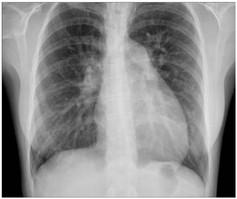
Fig. 1 Chest X-ray showed mild cardiomegaly and increased pulmonary vascularity in both lungs.
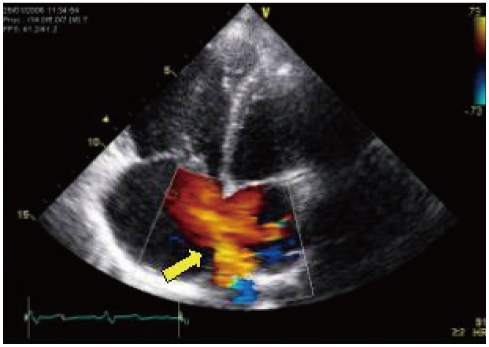
Fig. 2 Transthoracic echocardiography showed a large atrial septal defect (arrow), ostium secundum type (32-38 mm).
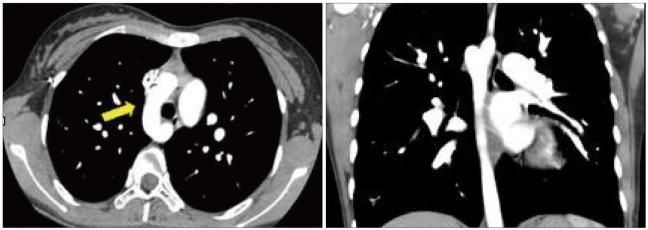
Fig. 3 Chest dynamic computed tomography showed right sided aortic arch (arrow).
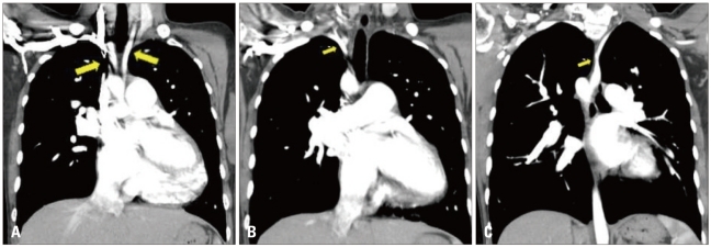
Fig. 4 Chest dynamic computed tomography showed the great vessels originated from the arch in the following order, left common carotid artery (right arrow) (A), right common carotid artery (left arrow) (A), right subclavian artery (B), and aberrant left subclavian artery (C). Left subclavian artery originated from descending aortic arch.
Reference
-
1. VanDyke CW, White RD. Congenital abnormalities of the thoracic aorta presenting in the adult. J Thorac Imaging. 1994; 9:230–245. PMID: 7830294.2. Shuford WH, Sybers RG. Shuford WH, Sybers RG, editors. The aortic arch and its malformations. Right aortic arch. 1974. Springfield: Charles C. Thomas;p. 52–68.3. Stewart JR, Kincaid OW, Titus JL. Right aortic arch: plain film diagnosis and significance. Am J Roentgenol Radium Ther Nucl Med. 1966; 97:377–389.4. Ruckman RN. Adams FH, Emmanouilides GC, Riemenschneider TA, editors. Anomalies of the aortic arch complex. Moss'heart disease in infants and adolescents. 1994. 5th ed. Baltimore: Williams & Wilkins.5. Stewart JR, Kincaid OW, Edwards JE. An atlas of vascular rings and related malformations of the aortic arch system. 1964. Springfield: Charles C. Thomas;p. 8–13. p. 124–219.6. Kaneda T, Lemura J, Zhang Z, Inoue T, Onoe M, Kitayama H, Nakamoto S, Oka H, Otaki M, Oku H. A case of Stanford type B aortic dissection involving a right-sided aortic arch with mirror-image branching and right-sided discending aorta. Thorac Cardiovasc Surg. 2001; 49:51–53. PMID: 11246740.7. Allen SR, Ignacio R, Falcone RA, Alonso MH, Brown RL, Garcia VF, Inge TH, Ryckman FC, Warner BW, Azizkhan RG, Tiao GM. The effect of a right-sided aortic arch on outcome in children with esophageal atresia and tracheoesophageal fistula. J Pediatr Surg. 2006; 41:479–483. PMID: 16516619.
- Full Text Links
-
- Actions
-
Cited
- CITED
-
- Close
- Share
-
- Similar articles
-
- A Case of Turner's Syndrome Associated with Atrial Septal Defect and Mitral Valve Prolapse
- A Case of Atrial Septal Defect in Identical Twins
- A Case of Interrupted Aortic Arch Diagnosed by Fetal Echocardiography
- A Case of Atrial Septal Aneurysm Associated with Atrial Septal Defect
- Distal type of aortopulmonary septal defect with aortic origin of right pulmonary artery and interruption of the aortic arch
