Ewha Med J.
2012 Mar;35(1):58-61. 10.12771/emj.2012.35.1.58.
Expansile Mass of the Rib
- Affiliations
-
- 1Department of Internal Medicine, St. Paul's Hospital, The Catholic University of Korea School of Medicine, Seoul, Korea. agmante@gmail.com
- 2Department of Radiology, St. Paul's Hospital, The Catholic University of Korea School of Medicine, Seoul, Korea.
- KMID: 2134948
- DOI: http://doi.org/10.12771/emj.2012.35.1.58
Abstract
- A 60-year-old man visited our hospital because of the incidentally found mass of the rib on chest radiography. Chest X-ray showed expansile bony hypertrophy on left 5th rib and bone setting of the computed tomography scan of chest revealed 4.2x2.5 cm sized, elongated bony expansion with geographic radiolucent lesion in the medullary cavity and cortical thinning. Technetium-99m bone scintigraphy showed diffusely increased radioactivity along the left 5th rib. We present this case to discuss about a possible differential diagnosis in this type of lesion.
Keyword
Figure
-

Fig. 1 Posteroanterior chest radiography shows expansile bony hypertrophy on left 5th rib.
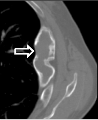
Fig. 2 Bone setting of the computed tomography scan of the chest shows elongated bony expansion with geographic radiolucent lesion in the medullary cavity and cortical thinning of left 5th rib (arrow).

Fig. 3 Technetium-99m bone scintigraphy shows diffusely increased uptake of isotope along the left 5th rib.
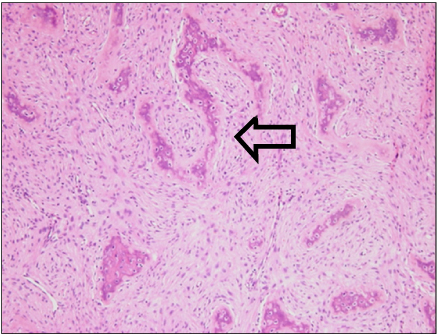
Fig. 4 Microscopic finding of excisional biopsy of the rib shows irregular spindles of woven bone (characteristic chinese letter, arrow) without osteoblastic rimming and a proliferation of fibroblast consistent with fibrous dysplasia (H&E stain, ×100).
Reference
-
1. Seo JS. Yoo HS, editor. Radiology of musculoskeletal system. Textbook of diagnostic radiology. 2003. 2nd ed. Seoul: Korea Medical Book Publishing;423–433.2. Baumgartner F, Section X. Bongard FS, Stomos MJ, Passaro E, editors. Cardiothoracic surgery. Surgery: a clinical approach. 1997. 1st ed. New York: Churchill Livingstone Inc.;375–379.3. Ryan MB, McMurtrey MJ, Roth JA. Current management of chest-wall tumors. Surg Clin North Am. 1989. 69:1061–1080.4. Daffner RH, Kirks DR, Gehweiler JA Jr, Heaston DK. Computed tomography of fibrous dysplasia. AJR Am J Roentgenol. 1982. 139:943–948.5. Riminucci M, Liu B, Corsi A, Shenker A, Spiegel AM, Robey PG, et al. The histopathology of fibrous dysplasia of bone in patients with activating mutations of the Gs alpha gene: site-specific patterns and recurrent histological hallmarks. J Pathol. 1999. 187:249–258.6. Hughes EK, James SL, Butt S, Davies AM, Saifuddin A. Benign primary tumours of the ribs. Clin Radiol. 2006. 61:314–322.7. DiCaprio MR, Enneking WF. Fibrous dysplasia. Pathophysiology, evaluation, and treatment. J Bone Joint Surg Am. 2005. 87:1848–1864.
