Evaluation of the effectiveness of the new tooth wear measurement parameters
- Affiliations
-
- 1Department of Oral Anatomy and Dental Research Institute, School of Dentistry, Seoul National University, Seoul, Korea. orana9@snu.ac.kr
- KMID: 2133269
- DOI: http://doi.org/10.5115/acb.2015.48.4.284
Abstract
- Nowadays, there has been an increasing interest in the preservation of natural dentition and the proper occlusion related to tooth wear for quality of life. To overcome the problems of the existing qualitative tooth wear analysis method, virtual three-dimensional models have been used. This study was designed to develop and validate a new quantitative method using tooth wear measurement parameters with angles obtained from virtual vectors and planes of the three-dimensional models. Sixteen parameters were evaluated in the virtual models of 20 students (7.57+/-1.55 years old) and 20 adults (56.85+/-6.34 years old). There were 12 angle and 4 height parameters, and the number of parameters measured from the virtual planes and vectors were 10 and 6, respectively. For each parameter, means and standard deviations were calculated, and an unpaired sample t test was performed to compare the young and the adult groups. Also, differences between the means were determined and expressed as percentages. The results were statistically significant between the two groups (P<0.001). In general, parameters using virtual vectors showed greater change than virtual plane. Although there were statistically significant differences among all parameters using virtual planes (P<0.001), the changes of the three angles were similar, except distolingual cusp angle. It was found that the parameters using virtual vectors were effective and tooth wear took place in both buccal and lingual cusps. Likewise, the validation of the new measurement parameters suggests that they can also be applied in the assessment of tooth wear related to dental biomaterials.
Keyword
MeSH Terms
Figure
-

Fig. 1 Reference points, vectors, and planes. (A) MBV1, DBV1, MLV1, DLV1. (B) MBV2, DBV2, MLV2, DLV2, BCV, LCV. (C) BP. (D) LP. MBV1 (mesiobuccal cusp vector 1), the vector connecting mesiobuccal cusp point (MBCP) and central pit point (CPP); DBV1 (distobuccal cusp vector 1), the vector connecting distobuccal cusp point (DBCP) and CPP; MLV1 (mesiolingual cusp vector 1), the vector connecting mesiolingual cusp point (MLCP) and CPP; DLV1 (distolingual cusp vector 1), the vector connecting distolingual cusp point (DLCP) and CPP; MBV2 (mesiobuccal cusp vector 2), the vector connecting MBCP and buccal lowest point (BLP); DBV2 (distobuccal cusp vector 2), the vector connecting DBCP and BLP; MLV2 (mesiolingual cusp vector 2), the vector connecting MLCP and lingual lowest point (LLP); DLV2 (distolingual cusp vector 2), the vector connecting DLCP and LLP; BCV (buccal cusps vector), the vector connecting MBCP and DBCP; LCV (lingual cusps vector), the vector connecting MLCP and DLCP; BP (buccal occlusal plane), the plane consisting of MBCP, DBCP, and MLCP; LP (lingual occlusal plane), the plane consisting of MBCP, MLCP, and DLCP.
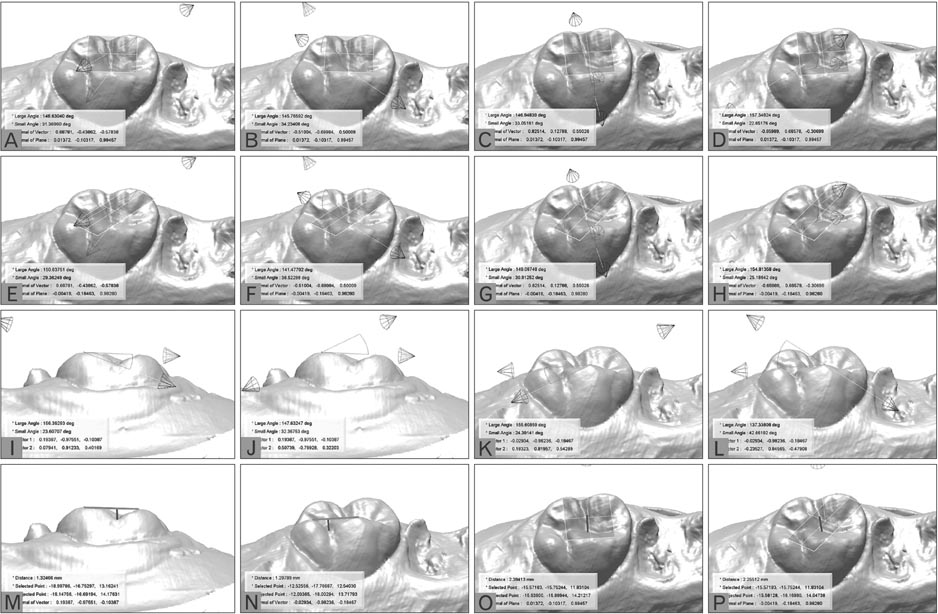
Fig. 2 Measurement parameters. (A) MBCA_BP. (B) DBCA_BP. (C) MLCA_BP. (D) DLCA_BP. (E) MBCA_LP. (F) DBCA_LP. (G) MLCA_LP. (H) DLCA_LP. (I) MBCA_BV. (J) DBCA_BV. (K) MLCA_LV. (L) DLCA_LV. (M) BCVH. (N) LCVH. (O) BCPH. (P) LCPH. MBCA_BP (mesiobuccal cusp angle with buccal occlusal plane [BP]), the angle between mesiobuccal cusp vector 1 (MBV1) and BP; DBCA_BP (distobuccal cusp angle with BP), the angle between distobuccal cusp vector 1 (DBV1) and BP; MLCA_BP (mesiolingual cusp angle with BP), the angle between mesiolingual cusp vector 1 (MLV1) and BP; DLCA_BP (distolingual cusp angle with BP), the angle between distolingual cusp vector 1 (DLV1) and BP; MBCA_LP (mesiobuccal cusp angle with lingual occlusal plane [LP]), the angle between MBV1 and LP; DBCA_LP (distobuccal cusp angle with LP), the angle between DBV1 and LP; MLCA_LP (mesiolingual cusp angle with LP), the angle between MLV1 and LP; DLCA_LP (distolingual cusp angle with LP), the angle between DLV1 and LP; MBCA_BV (mesiobuccal cusp angle with buccal cusps vector [BV]), the angle between mesiobuccal cusp vector 2 and BV; DBCA_BV (distobuccal cusp angle with BV), the angle between distobuccal cusp vector 2 and BV; MLCA_LV (mesiolingual cusp angle with lingual cusps vector [LV]), the angle between mesiolingual cusp vector 2 and LV; DLCA_LV (distolingual cusp angle with LV), the angle between distolingual cusp vector 2 and LV; BCVH (buccal cusps vector height), the shortest distance from buccal cusps vector to buccal lowest point; LCVH (lingual cusps vector height), the shortest distance from lingual cusps vector to lingual lowest point; BCPH (buccal cusps plane height), the shortest distance from BP to central pit point (CPP); LCPH (lingual cusps plane height), the shortest distance from LP to CPP.
Cited by 2 articles
-
Quantitative and qualitative evaluation on the accuracy of three intraoral scanners for human identification in forensic odontology
Eun-Jeong Bae, Eun-Jin Woo
Anat Cell Biol. 2022;55(1):72-78. doi: 10.5115/acb.21.136.Dental characteristics on panoramic radiographs as parameters for non-invasive age estimation: a pilot study
Harin Cheong, Akiko Kumagai, Sehyun Oh, Sang-Seob Lee
Anat Cell Biol. 2023;56(4):474-481. doi: 10.5115/acb.23.140.
Reference
-
1. Bishop K, Kelleher M, Briggs P, Joshi R. Wear now? An update on the etiology of tooth wear. Quintessence Int. 1997; 28:305–313.2. Smith BG. Toothwear: aetiology and diagnosis. Dent Update. 1989; 16:204–212.3. DeLong R. Intra-oral restorative materials wear: rethinking the current approaches: how to measure wear. Dent Mater. 2006; 22:702–711.4. Smith BG, Knight JK. An index for measuring the wear of teeth. Br Dent J. 1984; 156:435–438.5. Leinfelder KF, Taylor DF, Barkmeier WW, Goldberg AJ. Quantitative wear measurement of posterior composite resins. Dent Mater. 1986; 2:198–201.6. Magne P, Gallucci GO, Belser UC. Anatomic crown width/length ratios of unworn and worn maxillary teeth in white subjects. J Prosthet Dent. 2003; 89:453–461.7. Pigno MA, Hatch JP, Rodrigues-Garcia RC, Sakai S, Rugh JD. Severity, distribution, and correlates of occlusal tooth wear in a sample of Mexican-American and European-American adults. Int J Prosthodont. 2001; 14:65–70.8. Brosky ME, Major RJ, DeLong R, Hodges JS. Evaluation of dental arch reproduction using three-dimensional optical digitization. J Prosthet Dent. 2003; 90:434–440.9. DeLong R, Knorr S, Anderson GC, Hodges J, Pintado MR. Accuracy of contacts calculated from 3D images of occlusal surfaces. J Dent. 2007; 35:528–534.10. Lee JH, Paik KS, Chang MS, Lee SP. An evaluation of validity of measurements using digital caliper and three-dimensional virtual dental models. Korean J Anat. 2004; 37:209–218.11. Ha Y, Park YS, Nam SE, Lee JH, Lee JB, Lee SP. Development of quantitative tooth wear measurement parameters for Korean maxillary first molar. J Korea Res Soc Dent Mater. 2009; 36:157–166.12. Lee SP, Nam SE, Lee YM, Park YS, Hayashi K, Lee JB. The development of quantitative methods using virtual models for the measurement of tooth wear. Clin Anat. 2012; 25:347–358.13. Molnar S, Molnar IM. Dental arch shape and tooth wear variability. Am J Phys Anthropol. 1990; 82:385–395.14. Molnar S. Human tooth wear, tooth function and cultural variability. Am J Phys Anthropol. 1971; 34:175–189.15. Lovejoy CO. Dental wear in the Libben population: its functional pattern and role in the determination of adult skeletal age at death. Am J Phys Anthropol. 1985; 68:47–56.16. Molnar S, Richards L, McKee J, Molnar I. Tooth wear in Australian aboriginal populations from the River Murray Valley. Am J Phys Anthropol. 1989; 79:185–196.17. Johanson G. Age determinations from human teeth: a critical evaluation with special consideration of changes after fourteen years of age. Odontol Revy. 1971; 22:1–126.18. Molnar S, McKee JK, Molnar IM, Przybeck TR. Tooth wear rates among contemporary Australian Aborigines. J Dent Res. 1983; 62:562–565.19. Tomenchuk J, Mayhall JT. A correlation of tooth wear and age among modern Igloolik eskimos. Am J Phys Anthropol. 1979; 51:67–77.20. Hinton RJ. Form and patterning of anterior tooth wear among aboriginal human groups. Am J Phys Anthropol. 1981; 54:555–564.21. Mehta JD, Evans CC. A study of attrition of teeth in the Arkansas Indian skulls. Angle Orthod. 1966; 36:248–257.22. Mayhall JT, Kageyama I. A new, three-dimensional method for determining tooth wear. Am J Phys Anthropol. 1997; 103:463–469.23. Butler RJ. Age-related variability in occlusal wear planes. Am J Phys Anthropol. 1972; 36:381–390.24. Santini A, Land M, Raab GM. The accuracy of simple ordinal scoring of tooth attrition in age assessment. Forensic Sci Int. 1990; 48:175–184.25. Murphy TR. The timing and mechanism of the human masticatory stroke. Arch Oral Biol. 1965; 10:981–994.26. Woda A, Gourdon AM, Faraj M. Occlusal contacts and tooth wear. J Prosthet Dent. 1987; 57:85–93.27. Woda A, Vigneron P, Kay D. Nonfunctional and functional occlusal contacts: a review of the literature. J Prosthet Dent. 1979; 42:335–341.
