Korean J Orthod.
2015 Nov;45(6):333-340. 10.4041/kjod.2015.45.6.333.
Condylar hyperplasia: An updated review of the literature
- Affiliations
-
- 1Division of Oral and Maxillofacial Surgery, Department of Surgical Sciences, School of Dentistry, Marquette University, Milwaukee, WI, USA. luis.almeida@marquette.edu
- KMID: 2130595
- DOI: http://doi.org/10.4041/kjod.2015.45.6.333
Abstract
- Condylar hyperplasia (CH) is a rare disorder characterized by excessive bone growth that almost always presents unilaterally, resulting in facial asymmetry. Classification of the different types of CH can differ depending on the authors. Correct diagnosis is critical in determining the proper treatments and timing. This paper is a review of the recent literature on the epidemiology, etiology, diagnosis, classification, and surgical treatments of CH.
MeSH Terms
Figure
-
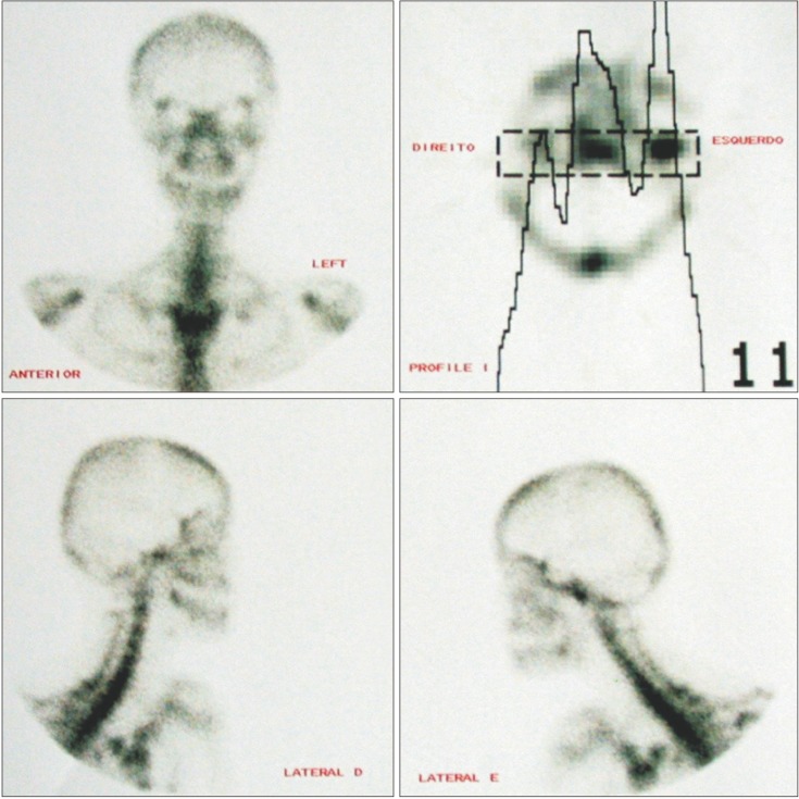
Figure 1 Scintigraphy showing activity in the left condyle.
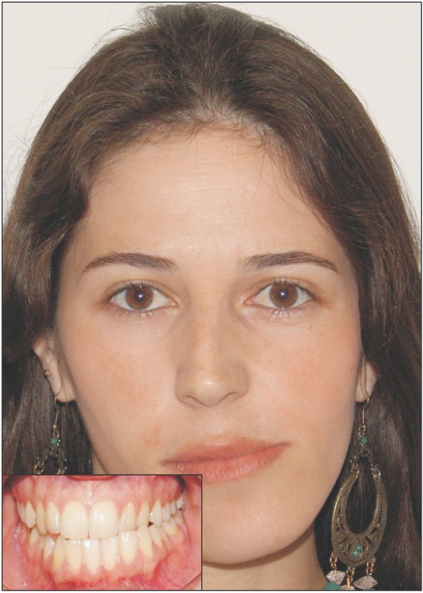
Figure 2 Clinical characteristics showing facial and occlusal deviation to the non-affected side.
Cited by 1 articles
-
Surgery-early approach combined with condylectomy for correction of severe facial asymmetry with mandibular condylar hyperplasia: a case report
Hikari Suzuki, Shinnosuke Nogami, Yoshio Otake, Yuri Takeda, Junji Sugawara, Tetsu Takahashi, Kensuke Yamauchi
J Korean Assoc Oral Maxillofac Surg. 2024;50(4):227-234. doi: 10.5125/jkaoms.2024.50.4.227.
Reference
-
2. Wolford LM, Movahed R, Perez DE. A classification system for conditions causing condylar hyperplasia. J Oral Maxillofac Surg. 2014; 72:567–595. PMID: 24388179.
Article3. Obwegeser HL, Makek MS. Hemimandibular hyperplasia--hemimandibular elongation. J Maxillofac Surg. 1986; 14:183–208. PMID: 3461097.
Article4. Nitzan DW, Katsnelson A, Bermanis I, Brin I, Casap N. The clinical characteristics of condylar hyperplasia: experience with 61 patients. J Oral Maxillofac Surg. 2008; 66:312–318. PMID: 18201615.
Article5. Raijmakers PG, Karssemakers LH, Tuinzing DB. Female predominance and effect of gender on unilateral condylar hyperplasia: a review and meta-analysis. J Oral Maxillofac Surg. 2012; 70:e72–e76. PMID: 21856058.
Article6. Hansson T, Oberg T, Carlsson GE, Kopp S. Thickness of the soft tissue layers and the articular disk in the temporomandibular joint. Acta Odontol Scand. 1977; 35:77–83. PMID: 266827.
Article7. Chen Y, Ke J, Long X, Meng Q, Deng M, Fang W, et al. Insulin-like growth factor-1 boosts the developing process of condylar hyperplasia by stimulating chondrocytes proliferation. Osteoarthritis Cartilage. 2012; 20:279–287. PMID: 22281262.
Article8. Renehan AG, Zwahlen M, Minder C, O'Dwyer ST, Shalet SM, Egger M. Insulin-like growth factor (IGF)-I, IGF binding protein-3, and cancer risk: systematic review and meta-regression analysis. Lancet. 2004; 363:1346–1353. PMID: 15110491.
Article9. Götz W, Lehmann TS, Appel TR, Rath-Deschner B, Dettmeyer R, Luder HU, et al. Distribution of insulin-like growth factors in condylar hyperplasia. Ann Anat. 2007; 189:347–349. PMID: 17695990.
Article10. Saridin CP, Raijmakers PG, Kloet RW, Tuinzing DB, Becking AG, Lammertsma AA. No signs of metabolic hyperactivity in patients with unilateral condylar hyperactivity: an in vivo positron emission tomography study. J Oral Maxillofac Surg. 2009; 67:576–581. PMID: 19231783.
Article11. Li QF, Rabie AB. A new approach to control condylar growth by regulating angiogenesis. Arch Oral Biol. 2007; 52:1009–1017. PMID: 17640614.
Article12. Pripatnanont P, Vittayakittipong P, Markmanee U, Thongmak S, Yipintsoi T. The use of SPECT to evaluate growth cessation of the mandible in unilateral condylar hyperplasia. Int J Oral Maxillofac Surg. 2005; 34:364–368. PMID: 16053843.
Article13. Saridin CP, Raijmakers PG, Tuinzing DB, Becking AG. Bone scintigraphy as a diagnostic method in unilateral hyperactivity of the mandibular condyles: a review and meta-analysis of the literature. Int J Oral Maxillofac Surg. 2011; 40:11–17. PMID: 20970961.
Article14. Lewis EL, Dolwick MF, Abramowicz S, Reeder SL. Contemporary imaging of the temporomandibular joint. Dent Clin North Am. 2008; 52:875–890. PMID: 18805233.
Article15. Laverick S, Bounds G, Wong WL. [18F]-fluoride positron emission tomography for imaging condylar hyperplasia. Br J Oral Maxillofac Surg. 2009; 47:196–199. PMID: 18926607.
Article16. Fariña RA, Becar M, Plaza C, Espinoza I, Franco ME. Correlation between single photon emission computed tomography, AgNOR count, and histomorphologic features in patients with active mandibular condylar hyperplasia. J Oral Maxillofac Surg. 2011; 69:356–361. PMID: 21122972.
Article17. Gray RJ, Sloan P, Quayle AA, Carter DH. Histopathological and scintigraphic features of condylar hyperplasia. Int J Oral Maxillofac Surg. 1990; 19:65–71. PMID: 2111360.
Article18. Bruce RA, Hayward JR. Condylar hyperplasia and mandibular asymmetry: a review. J Oral Surg. 1968; 26:281–290. PMID: 4867494.19. Wolford LM, Mehra P, Reiche-Fischel O, Morales-Ryan CA, García-Morales P. Efficacy of high condylectomy for management of condylar hyperplasia. Am J Orthod Dentofacial Orthop. 2002; 121:136–150. PMID: 11840126.
Article20. Slootweg PJ, Müller H. Condylar hyperplasia. A clinico-pathological analysis of 22 cases. J Maxillofac Surg. 1986; 14:209–214. PMID: 3461098.
Article21. Alyamani A, Abuzinada S. Management of patients with condylar hyperplasia: A diverse experience with 18 patients. Ann Maxillofac Surg. 2012; 2:17–23. PMID: 23483790.
Article22. Olate S, Netto HD, Rodriguez-Chessa J, Alister JP, de Albergaria-Barbosa J, de Moraes M. Mandible condylar hyperplasia: a review of diagnosis and treatment protocol. Int J Clin Exp Med. 2013; 6:727–737. PMID: 24179565.23. Saridin CP, Gilijamse M, Kuik DJ, te Veldhuis EC, Tuinzing DB, Lobbezoo F, et al. Evaluation of temporomandibular function after high partial condylectomy because of unilateral condylar hyperactivity. J Oral Maxillofac Surg. 2010; 68:1094–1099. PMID: 20149509.
Article24. Mehrotra D, Dhasmana S, Kamboj M, Gambhir G. Condylar hyperplasia and facial asymmetry: report of five cases. J Maxillofac Oral Surg. 2011; 10:50–56. PMID: 22379321.
Article25. Naini FB, Donaldson AN, McDonald F, Cobourne MT. Assessing the influence of asymmeftry affecting the mandible and chin point on perceived attractiveness in the orthognathic patient, clinician, and layperson. J Oral Maxillofac Surg. 2012; 70:192–206. PMID: 21571417.
Article26. Motamedi MH. Treatment of condylar hyperplasia of the mandible using unilateral ramus osteotomies. J Oral Maxillofac Surg. 1996; 54:1161–1169. PMID: 8859233.
Article27. Lippold C, Kruse-Losler B, Danesh G, Joos U, Meyer U. Treatment of hemimandibular hyperplasia: the biological basis of condylectomy. Br J Oral Maxillofac Surg. 2007; 45:353–360. PMID: 17145124.
Article28. Villanueva-Alcojol L, Monje F, González-García R. Hyperplasia of the mandibular condyle: clinical, histopathologic, and treatment considerations in a series of 36 patients. J Oral Maxillofac Surg. 2011; 69:447–455. PMID: 20828911.
Article29. Yamashita Y, Nakamura Y, Shimada T, Nomura Y, Hirashita A. Asymmetry of the lips of orthognathic surgery patients. Am J Orthod Dentofacial Orthop. 2009; 136:559–563. PMID: 19815159.
Article
- Full Text Links
-
- Actions
-
Cited
- CITED
-
- Close
- Share
-
- Similar articles
-
- Application and effects of condylectomy in asymmetric patients with condylar hyperplasia
- A Case Report Of Facial Asymmetry From Unilateral Condylar Hyperplasia
- Condylar Hyperplasia with Long-standing Temporomandibular Joint Dislocation
- Case report of the mandibular asymmetry with the unilateral condylar hyperplasia
- Correlation Between Mandibular Condylar Process Fracture and Temporomandibular Joint
