Comparison of prosthetic models produced by traditional and additive manufacturing methods
- Affiliations
-
- 1Department of Dental Laboratory, Science and Engineering, College of Health Science, Korea University, Republic of Korea. kuc2842@korea.ac.kr
- 2Department of Public Health Sciences, Graduate Shchool & BK21+Program in Public Health Sciences, Korea University, Republic of Korea.
- KMID: 2118265
- DOI: http://doi.org/10.4047/jap.2015.7.4.294
Abstract
- PURPOSE
The purpose of this study was to verify the clinical-feasibility of additive manufacturing by comparing the accuracy of four different manufacturing methods for metal coping: the conventional lost wax technique (CLWT); subtractive methods with wax blank milling (WBM); and two additive methods, multi jet modeling (MJM), and micro-stereolithography (Micro-SLA).
MATERIALS AND METHODS
Thirty study models were created using an acrylic model with the maxillary upper right canine, first premolar, and first molar teeth. Based on the scan files from a non-contact blue light scanner (Identica; Medit Co. Ltd., Seoul, Korea), thirty cores were produced using the WBM, MJM, and Micro-SLA methods, respectively, and another thirty frameworks were produced using the CLWT method. To measure the marginal and internal gap, the silicone replica method was adopted, and the silicone images obtained were evaluated using a digital microscope (KH-7700; Hirox, Tokyo, Japan) at 140X magnification. Analyses were performed using two-way analysis of variance (ANOVA) and Tukey post hoc test (alpha=.05).
RESULTS
The mean marginal gaps and internal gaps showed significant differences according to tooth type (P<.001 and P<.001, respectively) and manufacturing method (P<.037 and P<.001, respectively). Micro-SLA did not show any significant difference from CLWT regarding mean marginal gap compared to the WBM and MJM methods.
CONCLUSION
The mean values of gaps resulting from the four different manufacturing methods were within a clinically allowable range, and, thus, the clinical use of additive manufacturing methods is acceptable as an alternative to the traditional lost wax-technique and subtractive manufacturing.
Keyword
Figure
-

Fig. 1 Mimetic diagram of the 4 fabrication groups.
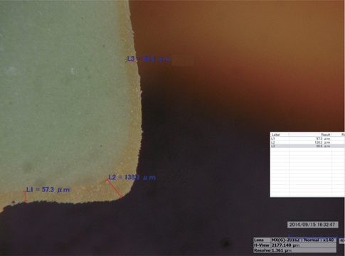
Fig. 2 An image of a model at 140× magnification using a digital microscope: L1; marginal gap, L2; rounded chamfer, L3; axial wall.
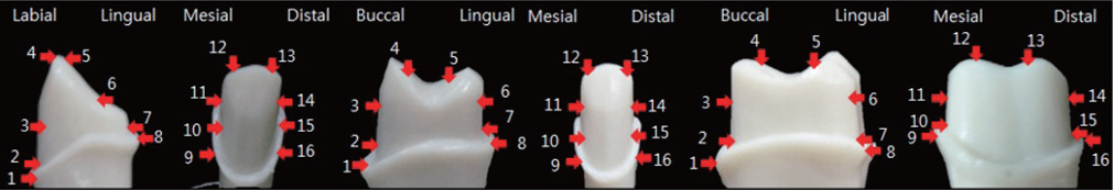
Fig. 3 The 16 measurement points for marginal and internal gap of crowns: marginal gap, (points 1, 8, 9, 16); rounded chamfer, (points 2, 7, 10, 15); axial wall, (points 3, 6, 11, 14); incisal or occlusal area, (points 4, 5, 12, 13).
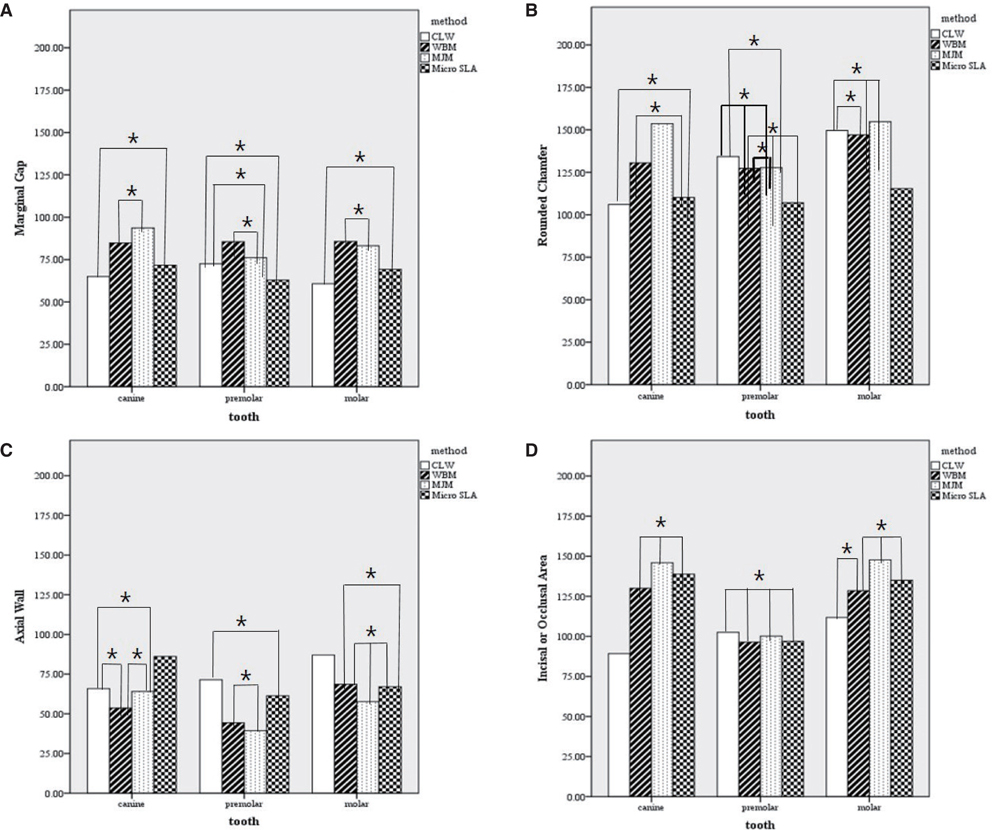
Fig. 4 Mean of MG, RC, AW, IA or OA according to the fabrication methods based on the tooth types. (A) mean values of marginal gap, (B) mean values of rounded chamfer, (C) mean values of axial wall, and (D) mean values of incisal or occlusal area.
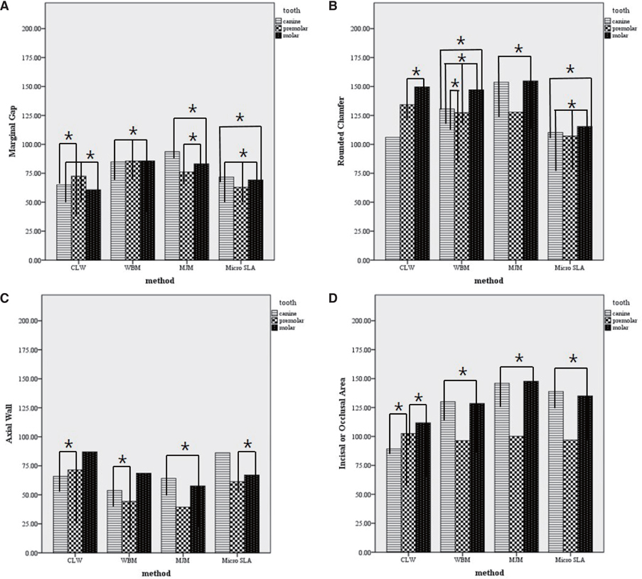
Fig. 5 Mean of MG, RC, AW, IA or OA according to the tooth types based on the fabrication methods. (A) Mean values of marginal gap, (B) Mean values of rounded chamfer, (C) Mean values of axial wall, and (D) Mean values of incisal or occlusal area.
Reference
-
1. Miyazaki T, Hotta Y, Kunii J, Kuriyama S, Tamaki Y. A review of dental CAD/CAM: current status and future perspectives from 20 years of experience. Dent Mater J. 2009; 28:44–56.2. Abduo J, Lyons K, Bennamoun M. Trends in computer-aided manufacturing in prosthodontics: a review of the available streams. Int J Dent. 2014; 2014:783948.3. Colpani JT, Borba M, Della Bona A. Evaluation of marginal and internal fit of ceramic crown copings. Dent Mater. 2013; 29:174–180.4. Anunmana C, Charoenchitt M, Asvanund C. Gap comparison between single crown and three-unit bridge zirconia substructures. J Adv Prosthodont. 2014; 6:253–258.5. Tamac E, Toksavul S, Toman M. Clinical marginal and internal adaptation of CAD/CAM milling, laser sintering, and cast metal ceramic crowns. J Prosthet Dent. 2014; 112:909–913.6. Almeida e Silva JS, Erdelt K, Edelhoff D, Araújo É, Stimmelmayr M, Vieira LC, Güth JF. Marginal and internal fit of four-unit zirconia fixed dental prostheses based on digital and conventional impression techniques. Clin Oral Investig. 2014; 18:515–523.7. Song TJ, Kwon TK, Yang JH, Han JS, Lee JB, Kim SH, Yeo IS. Marginal fit of anterior 3-unit fixed partial zirconia restorations using different CAD/CAM systems. J Adv Prosthodont. 2013; 5:219–225.8. Srikakula NK, Babu CS, Reddy JRK, Saiprasad SH, Raju ASR. Comparision of marginal fit of zirconium oxide copings generated using four different CAD-CAM systems-an in vitro study. J Res Adv Dent. 2014; 3:163–171.9. Sailer I, Fehér A, Filser F, Gauckler LJ, Lüthy H, Hämmerle CH. Five-year clinical results of zirconia frameworks for posterior fixed partial dentures. Int J Prosthodont. 2007; 20:383–388.10. Bornemann G, Lemelson S, Luthardt R. Innovative method for the analysis of the internal 3D fitting accuracy of Cerec-3 crowns. Int J Comput Dent. 2002; 5:177–182.11. Stanojevic M, Sljivic M, Plancak M, Djurdjevic D. Advanced investigation on rapid prototyping techniques in maxillofacial surgery and implanting preparation. J Technol Plast. 2014; 39:11–20.12. Snyder TJ, Andrews M, Weislogel M, Moeck P, Stone-Sundberg J, Birkes D, Hoffert MP, Lindeman A, Morrill J, Fercak O, Friedman S, Gunderson J, Ha A, McCollister J, Chen Y, Geile J, Wollman A, Attari B, Botnen N, Vuppuluri V, Shim J, Kaminsky W, Adams D, Graft J. 3D systems' technology overview and new applications in manufacturing, engineering, science, and education free access. 3D Pr Addit Manuf. 2014; 1:169–176.13. Sachs C, Groesser J, Stadelmann M, Schweiger J, Erdelt K, Beuer F. Full-arch prostheses from translucent zirconia: accuracy of fit. Dent Mater. 2014; 30:817–823.14. Quante K, Ludwig K, Kern M. Marginal and internal fit of metal-ceramic crowns fabricated with a new laser melting technology. Dent Mater. 2008; 24:1311–1315.15. Kim KB, Kim WC, Kim HY, Kim JH. An evaluation of marginal fit of three-unit fixed dental prostheses fabricated by direct metal laser sintering system. Dent Mater. 2013; 29:e91–e96.16. Örtorp A, Jönsson D, Mouhsen A, Vult von Steyern P. The fit of cobalt-chromium three-unit fixed dental prostheses fabricated with four different techniques: a comparative in vitro study. Dent Mater. 2011; 27:356–363.17. Kim KB, Kim JH, Kim WC, Kim JH. In vitro evaluation of marginal and internal adaptation of three-unit fixed dental prostheses produced by stereolithography. Dent Mater J. 2014; 33:504–509.18. Huang Z, Zhang L, Zhu J, Zhao Y, Zhang X. Clinical Marginal and Internal Fit of Crowns Fabricated Using Different CAD/CAM Technologies. J Prosthodont. 2015; 24:291–295.19. Kokubo Y, Ohkubo C, Tsumita M, Miyashita A, Vult von Steyern P, Fukushima S. Clinical marginal and internal gaps of Procera AllCeram crowns. J Oral Rehabil. 2005; 32:526–530.20. Nakamura T, Nonaka M, Maruyama T. In vitro fitting accuracy of copy-milled alumina cores and all-ceramic crowns. Int J Prosthodont. 2000; 13:189–193.21. Beuer F, Aggstaller H, Edelhoff D, Gernet W, Sorensen J. Marginal and internal fits of fixed dental prostheses zirconia retainers. Dent Mater. 2009; 25:94–102.22. Pelekanos S, Koumanou M, Koutayas SO, Zinelis S, Eliades G. Micro-CT evaluation of the marginal fit of different In-Ceram alumina copings. Eur J Esthet Dent. 2009; 4:278–292.23. Neves FD, Prado CJ, Prudente MS, Carneiro TA, Zancopé K, Davi LR, Mendonça G, Cooper LF, Soares CJ. Microcomputed tomography evaluation of marginal fit of lithium disilicate crowns fabricated by using chairside CAD/CAM systems or the heat-pressing technique. J Prosthet Dent. 2014; 112:1134–1140.24. Laurent M, Scheer P, Dejou J, Laborde G. Clinical evaluation of the marginal fit of cast crowns-validation of the silicone replica method. J Oral Rehabil. 2008; 35:116–122.25. Fransson B, Oilo G, Gjeitanger R. The fit of metal-ceramic crowns, a clinical study. Dent Mater. 1985; 1:197–199.26. McLean JW, von Fraunhofer JA. The estimation of cement film thickness by an in vivo technique. Br Dent J. 1971; 131:107–111.27. Belser UC, MacEntee MI, Richter WA. Fit of three porcelain-fused-to-metal marginal designs in vivo: a scanning electron microscope study. J Prosthet Dent. 1985; 53:24–29.28. Beuer F, Neumeier P, Naumann M. Marginal fit of 14-unit zirconia fixed dental prosthesis retainers. J Oral Rehabil. 2009; 36:142–149.29. Boening KW, Wolf BH, Schmidt AE, Kästner K, Walter MH. Clinical fit of Procera AllCeram crowns. J Prosthet Dent. 2000; 84:419–424.
- Full Text Links
-
- Actions
-
Cited
- CITED
-
- Close
- Share
-
- Similar articles
-
- Comparative evaluation of the subtractive and additive manufacturing on the color stability of fixed provisional prosthesis materials
- Fabrication of additive manufacturing interim denture and comparison with conventional interim denture: A case report
- A case of digital maxillary complete denture and mandibular implant overdenture fabricated by CAD-CAM technique
- Comparison of shear bond strength between various temporary prostheses resin blocks fabricated by subtractive and additive manufacturing methods bonded to self-curing reline resin
- On the marginal fidelity of all-ceramic core using CAD/CAM system
