J Adv Prosthodont.
2015 Apr;7(2):122-128. 10.4047/jap.2015.7.2.122.
Accuracy evaluation of metal copings fabricated by computer-aided milling and direct metal laser sintering systems
- Affiliations
-
- 1Department of Dental Laboratory Science and Engineering, College of Health Science, Korea University, Seoul, Republic of Korea. kjh2804@korea.ac.kr
- 2Department of Public Health Science, Graduate School & BK21+ Program in Public Health Science, Korea University, Seoul, Republic of Korea.
- KMID: 2118242
- DOI: http://doi.org/10.4047/jap.2015.7.2.122
Abstract
- PURPOSE
To assess the marginal and internal gaps of the copings fabricated by computer-aided milling and direct metal laser sintering (DMLS) systems in comparison to casting method.
MATERIALS AND METHODS
Ten metal copings were fabricated by casting, computer-aided milling, and DMLS. Seven mesiodistal and labiolingual positions were then measured, and each of these were divided into the categories; marginal gap (MG), cervical gap (CG), axial wall at internal gap (AG), and incisal edge at internal gap (IG). Evaluation was performed by a silicone replica technique. A digital microscope was used for measurement of silicone layer. Statistical analyses included one-way and repeated measure ANOVA to test the difference between the fabrication methods and categories of measured points (alpha=.05), respectively.
RESULTS
The mean gap differed significantly with fabrication methods (P<.001). Casting produced the narrowest gap in each of the four measured positions, whereas CG, AG, and IG proved narrower in computer-aided milling than in DMLS. Thus, with the exception of MG, all positions exhibited a significant difference between computer-aided milling and DMLS (P<.05).
CONCLUSION
Although the gap was found to vary with fabrication methods, the marginal and internal gaps of the copings fabricated by computer-aided milling and DMLS fell within the range of clinical acceptance (<120 microm). However, the statistically significant difference to conventional casting indicates that the gaps in computer-aided milling and DMLS fabricated restorations still need to be further reduced.
Keyword
MeSH Terms
Figure
-
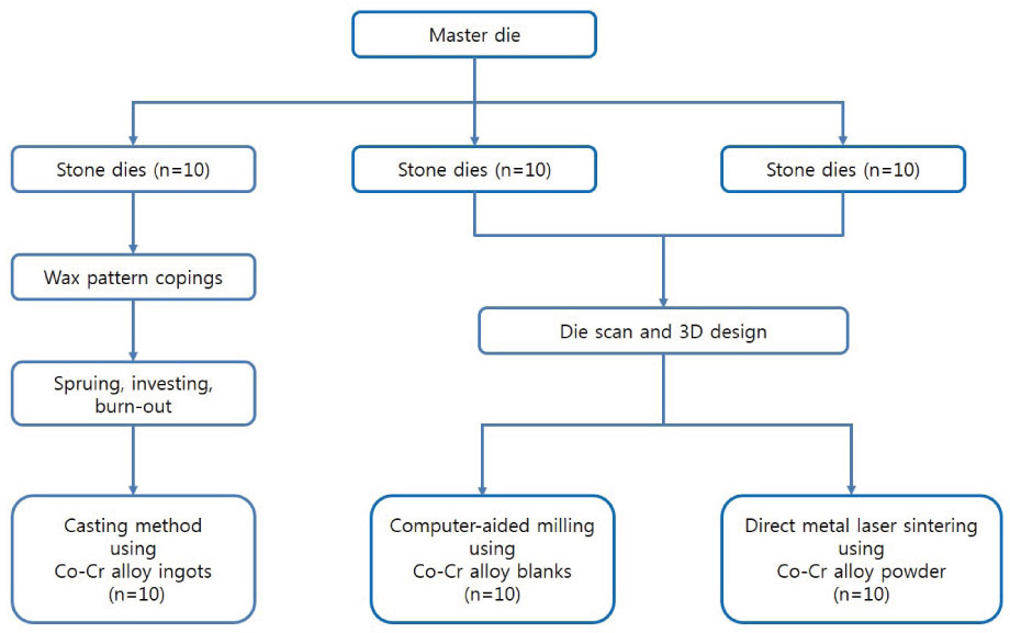
Fig. 1 Flowchart for the fabrication of metal copings.
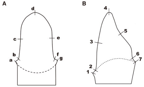
Fig. 2 Schematic diagram of the measuring points (A) mesiodistal section: a,g = marginal gap (MG), b,f = cervical gap (CG) (400 µm above MG), c,e = axial wall at internal gap (AG), d = incisal edge at internal gap (IG). (B) labiolingual section: 1,7 = MG, 2,6 = CG (400 µm above MG), 3,5 = AG, 4 = IG.

Fig. 3 Measurement of the marginal area silicone thickness by digital microscopy (magnification 160×).
Reference
-
1. Brecker SC. Porcelain baked to gold-A new medium in prosthodontics. J Prosthet Dent. 1956; 6:801–810.2. Johnston JF, Dykema RW, Cunningham DM. The use and construction of gold crowns with a fused porcelain veneer-A progress report. J Prosthet Dent. 1956; 6:811–821.3. Christensen GJ. Longevity of posterior tooth dental restorations. J Am Dent Assoc. 2005; 136:201–203.4. Miyazaki T, Hotta Y, Kunii J, Kuriyama S, Tamaki Y. A review of dental CAD/CAM: current status and future perspectives from 20 years of experience. Dent Mater J. 2009; 28:44–56.5. Weiss LE. Processes overview. In : Holdridge GM, Prinz FB, editors. Rapid Prototyping in Europe and Japan. Baltimore: Loyola College of Maryland;1997. p. 5–20.6. Petzold R, Zeilhofer HF, Kalender WA. Rapid protyping technology in medicine-basics and applications. Comput Med Imaging Graph. 1999; 23:277–284.7. Yadroitsev I, Bertrand Ph, Smurov I. Parametric analysis of the selective laser melting process. Appl Surf Sci. 2007; 253:8064–8069.8. Gardner FM. Margins of complete crowns-Literature review. J Prosthet Dent. 1982; 48:396–400.9. Wataha JC. Alloys for prosthodontic restorations. J Prosthet Dent. 2002; 87:351–363.10. Wataha JC, Messer RL. Casting alloys. Dent Clin North Am. 2004; 48:vii–viii. 499–512.11. Duncan JD. The casting accuracy of nickel-chromium alloys for fixed prostheses. J Prosthet Dent. 1982; 47:63–68.12. Ucar Y, Akova T, Akyil MS, Brantley WA. Internal fit evaluation of crowns prepared using a new dental crown fabrication technique: laser-sintered Co-Cr crowns. J Prosthet Dent. 2009; 102:253–259.13. Regish KM, Sharma D, Prithviraj DR, Nair A, Raghavan R. Evaluation and comparison of the internal fit and marginal accuracy of base metal (nickelchromium) and zirconia copings before and after ceramic veneering: a sem study. Eur J Prosthodont Restor Dent. 2013; 21:44–48.14. Eames WB, O'Neal SJ, Monteiro J, Miller C, Roan JD Jr, Cohen KS. Techniques to improve the seating of castings. J Am Dent Assoc. 1978; 96:432–437.15. Holmes JR, Bayne SC, Holland GA, Sulik WD. Considerations in measurement of marginal fit. J Prosthet Dent. 1989; 62:405–408.16. van Noort R. The future of dental devices is digital. Dent Mater. 2012; 28:3–12.17. Beuer F, Schweiger J, Edelhoff D. Digital dentistry: an overview of recent developments for CAD/CAM generated restorations. Br Dent J. 2008; 204:505–511.18. Örtorp A, Jönsson D, Mouhsen A, Vult von Steyern P. The fit of cobalt-chromium three-unit fixed dental prostheses fabricated with four different techniques: a comparative in vitro study. Dent Mater. 2011; 27:356–363.19. Quante K, Ludwig K, Kern M. Marginal and internal fit of metal-ceramic crowns fabricated with a new laser melting technology. Dent Mater. 2008; 24:1311–1315.20. Beuer F, Aggstaller H, Edelhoff D, Gernet W, Sorensen J. Marginal and internal fits of fixed dental prostheses zirconia retainers. Dent Mater. 2009; 25:94–102.21. Laurent M, Scheer P, Dejou J, Laborde G. Clinical evaluation of the marginal fit of cast crowns-validation of the silicone replica method. J Oral Rehabil. 2008; 35:116–122.22. Sorensen JA. A standardized method for determination of crown margin fidelity. J Prosthet Dent. 1990; 64:18–24.23. Pelekanos S, Koumanou M, Koutayas SO, Zinelis S, Eliades G. Micro-CT evaluation of the marginal fit of different In-Ceram alumina copings. Eur J Esthet Dent. 2009; 4:278–292.24. Rahme HY, Tehini GE, Adib SM, Ardo AS, Rifai KT. In vitro evaluation of the "replica technique" in the measurement of the fit of Procera crowns. J Contemp Dent Pract. 2008; 9:25–32.25. Council on Dental Materials and Devices. Revised American National Standards Instititute/American Dental Association Specification No. 8 for Zinc Phosphate Cement. J Am Dent Assoc. 1978; 96:121–123.26. Assif D, Rimer Y, Aviv I. The flow of zinc phosphate cement under a full-coverage restoration and its effect on marginal adaptation according to the location of cement application. Quintessence Int. 1987; 18:765–774.27. Hung SH, Hung KS, Eick JD, Chappell RP. Marginal fit of porcelain-fused-to-metal and two types of ceramic crown. J Prosthet Dent. 1990; 63:26–31.28. Gulker I. Margins. N Y State Dent J. 1985; 51:213–215. 21729. McLean JW, von Fraunhofer JA. The estimation of cement film thickness by an in vivo technique. Br Dent J. 1971; 131:107–111.
- Full Text Links
-
- Actions
-
Cited
- CITED
-
- Close
- Share
-
- Similar articles
-
- Evaluation of marginal discrepancy in metal frameworks fabricated by sintering-based computer-aided manufacturing methods
- Fabricating a Ceramic-Pressed-to-Metal Restoration with Computer-Aided Design, Computer-Aided Manufacturing and Selective Laser Sintering: A Case Report
- Fixed prostheses fabricated by direct metal laser sintering system: case report
- Influence of finish line design on the marginal fit of nonprecious metal alloy coping fabricated by 3D printing, milling and casting using CAD-CAM
- Influence of the accuracy of abutment tooth preparation on the marginal adaptation of Co-Cr alloy copings fabricated with a selective laser sintering technology
