J Adv Prosthodont.
2013 Aug;5(3):262-269. 10.4047/jap.2013.5.3.262.
Perceived color shift of ceramics according to the change of illuminating light with spectroradiometer
- Affiliations
-
- 1Department of Dentistry, College of Medicine, University of Ulsan, Asan Medical Center, Seoul, Republic of Korea.
- 2State Key Laboratory of Multiphase Complex Systems, Institute of Process Engineering, Chinese Academy of Sciences, Beijing, China.
- 3Institute for Clinical Performance of Biomaterials (ICPB), Seoul, Republic of Korea. ykleedm@gmail.com
- KMID: 2118165
- DOI: http://doi.org/10.4047/jap.2013.5.3.262
Abstract
- PURPOSE
Perceived color of ceramics changes by the spectral power distribution of ambient light. This study aimed to quantify the amount of shifts in color and color coordinates of clinically simulated seven all-ceramics due to the switch of three ambient light sources using a human vision simulating spectroradiometer.
MATERIALS AND METHODS
CIE color coordinates, such as L*, a* and b*,of ceramic specimens were measured under three light sources, which simulate the CIE standard illuminant D65 (daylight), A (incandescent lamp), and F9 (fluorescent lamp). Shifts in color and color coordinate by the switch of lights were determined. Influence of the switched light (D65 to A, or D65 to F9), shade of veneer ceramics (A2 or A3), and brand of ceramics on the shifts was analyzed by a three-way ANOVA.
RESULTS
Shifts in color and color coordinates were influenced by three factors (P<.05). Color shifts by the switch to A were in the range of 5.9 to 7.7 DeltaE*abunits, and those by the switch to F9 were 7.7 to 10.2; all of which were unacceptable (DeltaE*ab > 5.5). When switched to A, CIE a* increased (Deltaa*: 5.6 to 7.6), however, CIE b* increased (Deltab*: 4.9 to 7.8) when switched to F9.
CONCLUSION
Clinically simulated ceramics demonstrated clinically unacceptable color shifts according to the switches in ambient lights based on spectroradiometric readings. Therefore, shade matching and compatibility evaluation should be performed considering ambient lighting conditions and should be done under most relevant lighting condition.
Figure
-

Fig. 1 Vectorial shifts of lightness (CIE L*) and chroma for A2-veneered ceramics by switch of lights.
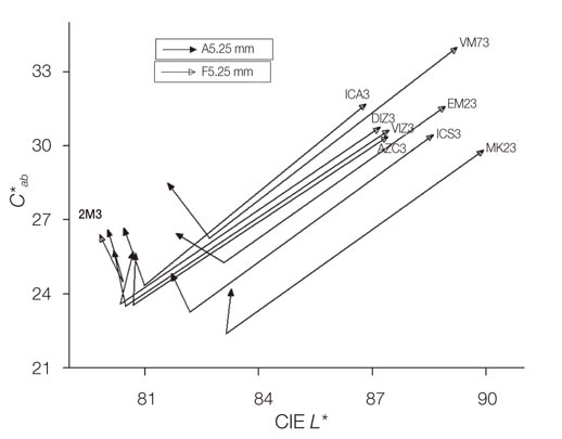
Fig. 2 Vectorial shifts of lightness (CIE L*) and chroma for A3-veneered ceramics by switch of lights.
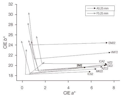
Fig. 3 Vectorial shifts of CIE a* and b* values for A2-veneered ceramics by switch of lights.
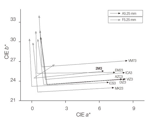
Fig. 4 Vectorial shifts of CIE a* and b* for A3-veneered ceramics by switch of lights.
Reference
-
1. Vichi A, Louca C, Corciolani G, Ferrari M. Color related to ceramic and zirconia restorations: a review. Dent Mater. 2011; 27:97–108.2. Barão VA, Gennari-Filho H, Goiato MC, dos Santos DM, Pesqueira AA. Factors to achieve aesthetics in all-ceramic restorations. J Craniofac Surg. 2010; 21:2007–2012.3. Lee YK, Yu B, Lim JI, Lim HN. Perceived color shift of a shade guide according to the change of illuminant. J Prosthet Dent. 2011; 105:91–99.4. Rosenstiel SF, Land MF, Fujimoto J. Contemporary fixed prothodontics. 4th ed. St. Louis: Mosby;2006. p. 774.5. Anusavice K. Philips' science of dental materials. 11th ed. St. Louis: Saunders;2003. p. 655–719.6. Zhang Y, Kim JW. Graded zirconia glass for resistance to veneer fracture. J Dent Res. 2010; 89:1057–1062.7. Barnes D, Gingell JC, George D, Adachi E, Jefferies S, Sundar VV. Clinical evaluation of an all-ceramic restorative system: a 36-month clinical evaluation. Am J Dent. 2010; 23:87–92.8. Park JH, Lee YK, Lim BS. Influence of illuminants on the color distribution of shade guides. J Prosthet Dent. 2006; 96:402–411.9. Judeh A, Al-Wahadni A. A comparison between conventional visual and spectrophotometric methods for shade selection. Quintessence Int. 2009; 40:e69–e79.10. Lehmann KM, Igiel C, Schmidtmann I, Scheller H. Four color-measuring devices compared with a spectrophotometric reference system. J Dent. 2010; 38:Suppl 2. e65–e70.11. Ishikawa-Nagai S, Yoshida A, Da Silva JD, Miller L. Spectrophotometric analysis of tooth color reproduction on anterior all-ceramic crowns: Part 1: analysis and interpretation of tooth color. J Esthet Restor Dent. 2010; 22:42–52.12. Johnston WM, Hesse NS, Davis BK, Seghi RR. Analysis of edge-losses in reflectance measurements of pigmented maxillofacial elastomer. J Dent Res. 1996; 75:752–760.13. Yap AU, Sim CP, Loh WL, Teo JH. Human-eye versus computerized color matching. Oper Dent. 1999; 24:358–363.14. Wyszecki G, Stiles WS. Color science - Concepts and methods, quantitative data and formulae. 2nd ed. New York: John Wiley and Sons;2000.15. Marcus RT. The measurement of color. In : Nassau K, editor. Color for science, art and technology. Amsterdam: Elsevier;1998. p. 48–49.16. Lee YK, Lim BS, Kim CW, Powers JM. Color characteristics of low-chroma and high-translucence dental resin composites by different measuring modes. J Biomed Mater Res. 2001; 58:613–621.17. Lee YK, Powers JM. Color difference of four esthetic restorative materials by the illuminant. Am J Dent. 2005; 18:359–363.18. Yu B, Lee YK. Color difference of all-ceramic materials by the change of illuminants. Am J Dent. 2009; 22:73–78.19. van der Burgt TP, ten Bosch JJ, Borsboom PC, Plasschaert AJ. A new method for matching tooth colors with color standards. J Dent Res. 1985; 64:837–841.20. Lee YK, Yoon TH, Lim BS, Kim CW, Powers JM. Effects of colour measuring mode and light source on the colour of shade guides. J Oral Rehabil. 2002; 29:1099–1107.21. Kim SH, Lee YK, Lim BS, Rhee SH, Yang HC. Metameric effect between dental porcelain and porcelain repairing resin composite. Dent Mater. 2007; 23:374–379.22. Volpato CA, Monteiro S Jr, de Andrada MC, Fredel MC, Petter CO. Optical influence of the type of illuminant, substrates and thickness of ceramic materials. Dent Mater. 2009; 25:87–93.23. Corcodel N, Helling S, Rammelsberg P, Hassel AJ. Metameric effect between natural teeth and the shade tabs of a shade guide. Eur J Oral Sci. 2010; 118:311–316.24. Cha HS, Lee YK. Difference in illuminant-dependent color changes of shade guide tabs by the shade designation relative to three illuminants. Am J Dent. 2009; 22:350–356.25. Gokce HS, Piskin B, Ceyhan D, Gokce SM, Arisan V. Shade matching performance of normal and color vision-deficient dental professionals with standard daylight and tungsten illuminants. J Prosthet Dent. 2010; 103:139–147.26. Carsten DL. Successful shade matching--what does it take. Compend Contin Educ Dent. 2003; 24:175–182.27. Commission Internationale de l'Eclairage (CIE) Colorimetry - technical report. CIE Pub. No. 15. 3rd ed. Vienna: Bureau Central de la CIE;2004.28. Lee YK, Yu B, Lee SH, Cho MS, Lee CY, Lim HN. Variation in instrument-based color coordinates of esthetic restorative materials by measurement method-A review. Dent Mater. 2010; 26:1098–1105.29. van der Burgt TP, ten Bosch JJ, Borsboom PC, Kortsmit WJ. A comparison of new and conventional methods for quantification of tooth color. J Prosthet Dent. 1990; 63:155–162.30. Bolt RA, Bosch JJ, Coops JC. Influence of window size in small-window color measurements, particularly of teeth. Phys Med Biol. 1994; 39:1133–1142.31. Lee YK, Cha HS, Ahn JS. Layered color of all-ceramic core and veneer ceramics. J Prosthet Dent. 2007; 97:279–286.32. Gozalo-Diaz D, Johnston WM, Wee AG. Estimating the color of maxillary central incisors based on age and gender. J Prosthet Dent. 2008; 100:93–98.33. Bayindir F, Bayindir YZ, Gozalo-Diaz DJ, Wee AG. Coverage error of gingival shade guide systems in measuring color of attached anterior gingiva. J Prosthet Dent. 2009; 101:46–53.34. Li Q, Yu H, Wang YN. In vivo spectroradiometric evaluation of colour matching errors among five shade guides. J Oral Rehabil. 2009; 36:65–70.35. Ghinea R, Pérez MM, Herrera LJ, Rivas MJ, Yebra A, Paravina RD. Color difference thresholds in dental ceramics. J Dent. 2010; 38:Suppl 2. e57–e64.36. Douglas RD, Steinhauer TJ, Wee AG. Intraoral determination of the tolerance of dentists for perceptibility and acceptability of shade mismatch. J Prosthet Dent. 2007; 97:200–208.37. Davis S. Color perception - philosophical, psychological, artistic, and cognitive perspectives. New York: Oxford University Press;2000. p. 88–90.38. O'Brien WJ, Groh CL, Boenke KM. One-dimensional color order system for dental shade guides. Dent Mater. 1989; 5:371–374.39. Ferreira D, Monard LA. Measurement of spectral reflectance and colorimetric properties of Vita shade guides. J Dent Assoc S Afr. 1991; 46:63–65.40. Lee YK, Powers JM. Metameric effect between resin composite and dentin. Dent Mater. 2005; 21:971–976.41. Heffernan MJ, Aquilino SA, Diaz-Arnold AM, Haselton DR, Stanford CM, Vargas MA. Relative translucency of six all-ceramic systems. Part I: core materials. J Prosthet Dent. 2002; 88:4–9.42. Paravina RD, Westland S, Kimura M, Powers JM, Imai FH. Color interaction of dental materials: blending effect of layered composites. Dent Mater. 2006; 22:903–908.43. Lee YK, Yu B, Lim HN. Lightness, chroma, and hue distributions of a shade guide as measured by a spectroradiometer. J Prosthet Dent. 2010; 104:173–181.44. Kim JG, Yu B, Lee YK. Correlations between color differences based on three color-difference formulas using dental shade guide tabs. J Prosthodont. 2009; 18:135–140.
- Full Text Links
-
- Actions
-
Cited
- CITED
-
- Close
- Share
-
- Similar articles
-
- The effect of repeated firings on the color change and surface roughness of dental ceramics
- The effect of repeated firings on the color change of dental ceramics using different glazing methods
- Influences of luting cement shade on the color of various translucent monolithic zirconia and lithium disilicate ceramics for veneer restorations
- Effect of surface finishing treatments on the color stability of CAD/CAM materials
- Spectrophotometic analysis of the influence of substrate on the color of dental ceramics
