J Korean Ophthalmol Soc.
2009 Mar;50(3):471-476. 10.3341/jkos.2009.50.3.471.
Various Treatments Using Invaluable Donor Cornea
- Affiliations
-
- 1Department of Ophthalmology, Chosun University College of Medicine, Gwanju, Korea. clearcornea@paran.com
- KMID: 2111261
- DOI: http://doi.org/10.3341/jkos.2009.50.3.471
Abstract
-
PURPOSE: To report cases of transplanting a donor's 2 corneas to 5 patients suffering from several corneal diseases.
CASE SUMMARY
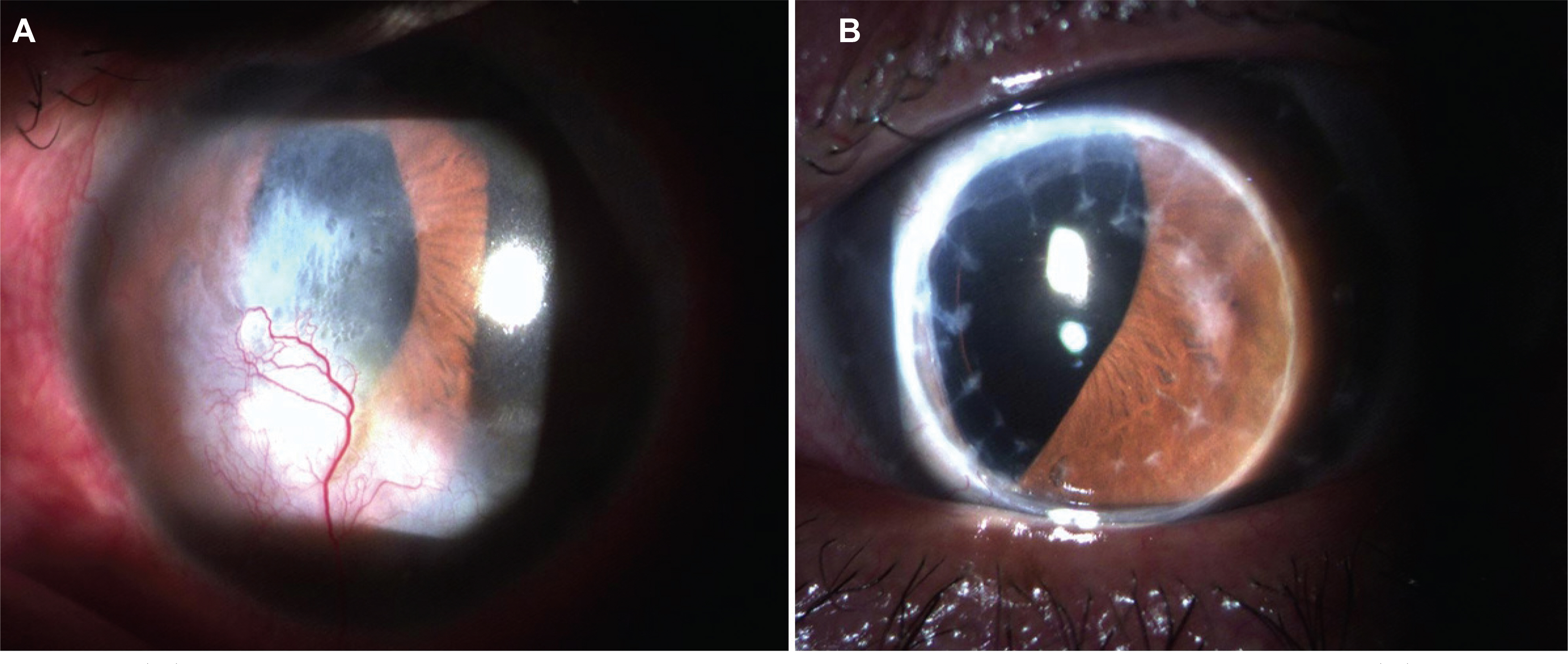
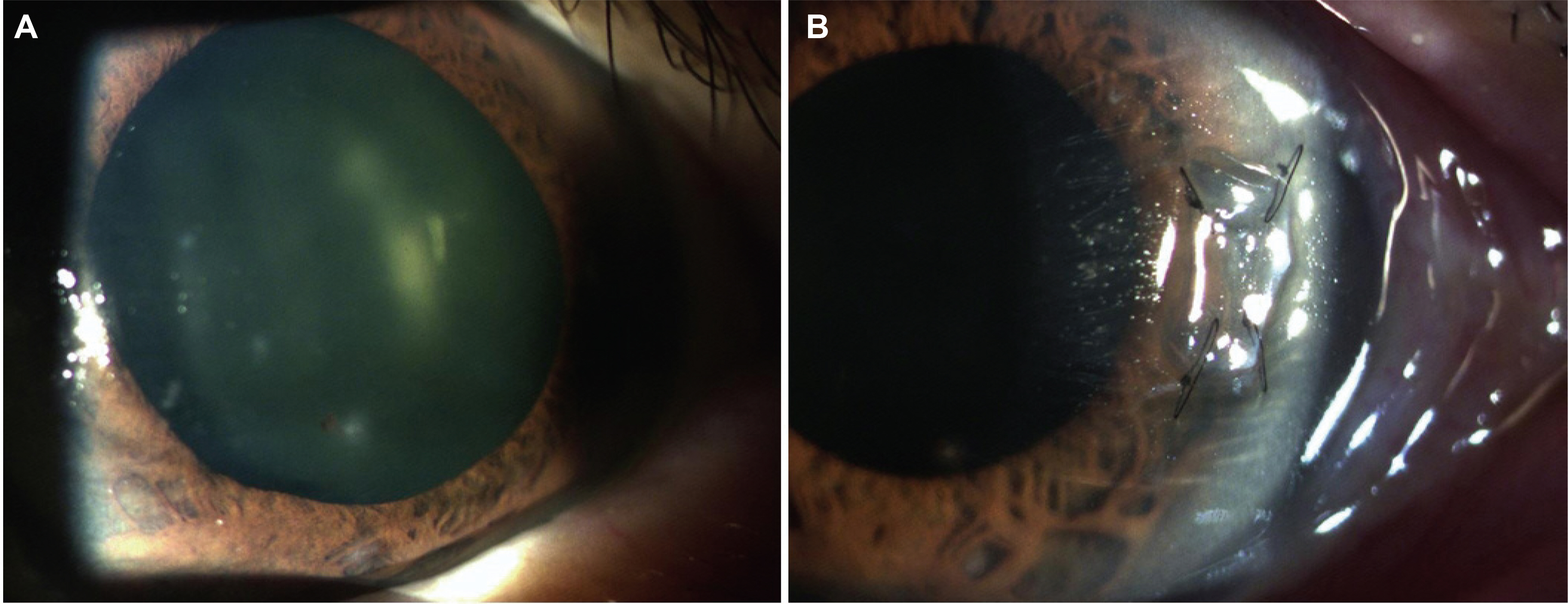
Two corneas were donated from a 66-year-old donor, who suffered from brain damage due to asphyxia, one hour after being pronounced dead by doctors. Two penetrating keratoplasties and 3 partial lamellar keratoplasties were performed for patients with corneal opacity, corneal ulcer and corneal perforation. After the procedure all grafts were stable.
CONCLUSIONS
Under the present circumstances of decreasing donations of corneas after death and the increasing demand for keratoplasty in Korea, the mutual cooperation among hospitals to treat more than one patient using one donated cornea is a method the authors believe can alleviate this situation.
MeSH Terms
Figure
Cited by 1 articles
-
Four Cases of Split Cornea Transplantation from a Single Cornea
Hyo Won Kim, Ho Sik Hwang, Sung A Lim, Man Soo Kim
J Korean Ophthalmol Soc. 2016;57(6):988-993. doi: 10.3341/jkos.2016.57.6.988.
Reference
-
References
1. Barron BA, Penetrating keratoplasty. The Cornea. 2nd ed.New York: Churchill Livingstone Inc;1998. p. 805–10.2. Choi SH, Lee YW, Kim HM, et al. Epidemiologic studies of keratoplasty in Korea. J Korean Ophthalmol Soc. 2006; 47:538–547.3. Terry MA, Ousley PJ. Small incision deep lamella endothelial keratoplasty. six-month results in the first prospective clinical study. Cornea. 2005; 24:59–65.4. Melles GR, Eggink FA, Launder F, et al. A surgical technique for posterior lamella keratoplasty. Cornea. 1998; 17:618–25.5. Terry MA, Ousley PJ. Endothelial replacement without surface corneal incisions or sutures. Cornea. 2001; 20:14–8.
Article6. Terry MA, Ousley PJ. Deep lamella endothelial keratoplasty in the first United States patients. Cornea. 2001; 20:239–43.7. Uhm SL, Chung SK, Myung YW, Rhee SW. Clinical analysis of keratoplasty over a 23 year-time span. J Korean Ophthalmol Soc. 1991; 32:421–9.8. Ha DW, Kim CK, Lee SE, et al. Penetrating keratoplasty results in 275 cases. J Korean Ophthalmol Soc. 2001; 42:20–29.9. Eye bank association of America. 2002 eye banking statistical report. Washington, DC: Eye Bank Association of America;2003.10. Means TL, Geroski DH, L'Hernault N, et al. The corneal epithelium after Optisol-GS storage. Cornea. 1996; 15:599–605.
Article11. Vajpayee RB, Sharma N, Jhanji V, et al. One donor cornea for 3 recipients. Arch Ophthalmol. 2007; 125:552–4.
Article
- Full Text Links
- Actions
-
Cited
- CITED
-
- Close
- Share
- Similar articles
-
- The Clinical Evaluations of the Penetrating Keratoplasty with Imported Donor Corneas
- Development of an Instrument for Slit-lamp Examination of Donor Corneas in Preservation Medium
- Lamellar Keratopasty
- Tectonic Deep Anterior Lamellar Keratoplasty in Impending Corneal Perforation Using Cryopreserved Cornea
- A Case of Therapeutic Keratoplasty Using Cryo-preservative Cornea in Candida albicans Keratitis


