J Korean Orthop Assoc.
2012 Jun;47(3):222-226. 10.4055/jkoa.2012.47.3.222.
Periprosthetic Fracture after Locked Plating in the Osteoporotic Long Bone Fracture
- Affiliations
-
- 1Department of Orthopedic Surgery, Guri Hospital, Hanyang University College of Medicine, Guri, Korea. kcpark@hanyang.ac.kr
- KMID: 2106652
- DOI: http://doi.org/10.4055/jkoa.2012.47.3.222
Abstract
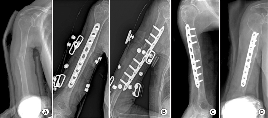
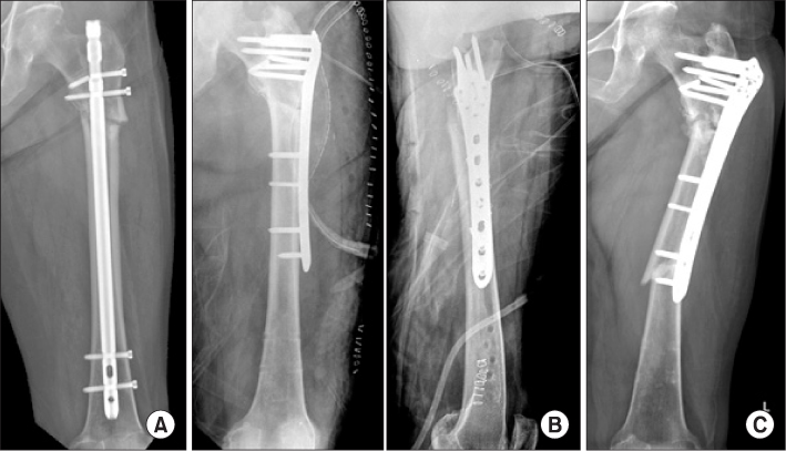
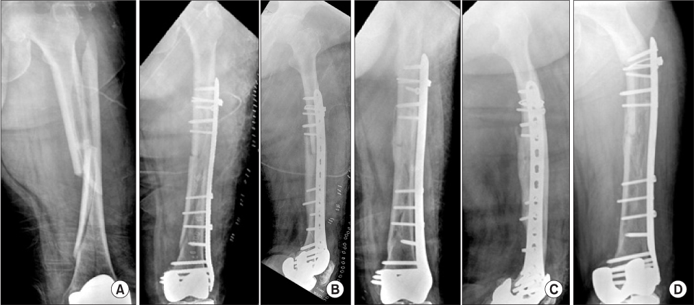
- The locking plate is used for osteoporotic long bone fracture because of the ability to affect a lock between the plate and locking screws that prevents reduction loss, angular deformity and axial deformity. However, it has been reported that locked plating can increase the periprosthetic fracture risk in osteoporotic bone compared with conventional plating. We report 3 cases of periprosthetic fracture after the use of locked plating in osteoporotic long bone fracture and a review of the relevant literatures.
Keyword
Figure
Cited by 1 articles
-
Comparison of Locking versus Dynamic Compression Plates for Treatment of Diaphyseal Forearm Fracture
Yong Chan Lee, Hong Je Kang
J Korean Soc Surg Hand. 2015;20(4):168-175. doi: 10.12790/jkssh.2015.20.4.168.
Reference
-
1. Frigg R. Development of the locking compression plate. Injury. 2003. 34:Suppl 2. B6–B10.
Article2. Sommer C, Gautier E, Müller M, Helfet DL, Wagner M. First clinical results of the locking compression plate (LCP). Injury. 2003. 34:Suppl 2. B43–B54.
Article3. Bottlang M, Doornink J, Byrd GD, Fitzpatrick DC, Madey SM. A nonlocking end screw can decrease fracture risk caused by locked plating in the osteoporotic diaphysis. J Bone Joint Surg Am. 2009. 91:620–627.
Article4. Fulkerson E, Egol KA, Kubiak EN, Liporace F, Kummer FJ, Koval KJ. Fixation of diaphyseal fractures with a segmental defect: a biomechanical comparison of locked and conventional plating techniques. J Trauma. 2006. 60:830–835.
Article5. Haidukewych GJ. Innovations in locking plate technology. J Am Acad Orthop Surg. 2004. 12:205–212.
Article6. Sim JC, Chung NS, Hong KD, Ha SS, Kang JH. Treatment of fractures of the distal radius using locking compression plate. J Korean Fract Soc. 2005. 18:100–104.
Article7. Davenport SR, Lindsey RW, Leggon R, Miclau T, Panjabi M. Dynamic compression plate fixation: a biomechanical comparison of unicortical vs bicortical distal screw fixation. J Orthop Trauma. 1988. 2:146–150.
Article
- Full Text Links
- Actions
-
Cited
- CITED
-
- Close
- Share
- Similar articles
-
- Locked Plating in Elderly Patients with Distal Femur Fracture: How to Avoid Complications?
- Plate Designed for Wiring ( CHO Plate )
- Volar Plating of Distal Radius Fractures
- The Outcome and Complications of the Locked Plating Management for the Periprosthetic Distal Femur Fractures after a Total Knee Arthroplasty
- Bone Density and Mechanical Property after Locked Intramedullary Nailing of the Long Bone Fractures: Preliminary Study of QCT & Lunar of 15 Long Bone Fractures


