J Korean Soc Endocrinol.
2005 Feb;20(1):84-89. 10.3803/jkes.2005.20.1.84.
A Case of Primary Squamous Cell Carcinoma of the Thyroid Gland
- Affiliations
-
- 1Department of Internal Medicine, Inje University College of Medicine, Paik Hospital, Pusan, Korea.
- 2Department of Pathology, Inje University College of Medicine, Paik Hospital, Pusan, Korea.
- KMID: 2100457
- DOI: http://doi.org/10.3803/jkes.2005.20.1.84
Abstract
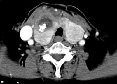
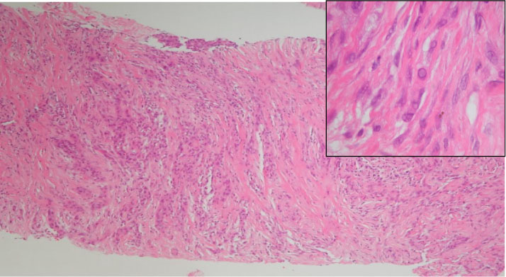
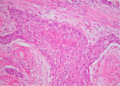
- Primary squamous cell carcinoma of the thyroid gland is an extremely rare case to observe and represents less than 1% in all the primary thyroid malignancies. Normally, squamous epithelium is absent in the thyroid gland and presently; its origin is believed to arise from metaplasia of follicular epithelium. Cancer has very aggressive clinical behavior and a very poor prognosis with survival rates of less than 1 year. The best chances of survival have been achieved with complete resection followed by postoperative radiotherapy. Recently, we came across a case of 80-year-old woman with primary squamous cell cacinoma of the thyroid gland present in the background of Hashimoto's thyroiditis. The patient had swelling in the anterior neck portion from the past 20 days. On physical examinaton, 3x3cm2 hard and fixed ill defined mass was detected in the right lobe of thyroid. Repeated fine needle aspiration biopsy of the thyroid revealed the presence of carcinoma. Apparently, Palliative thyroidectomy was performed after 3 months of diagnosis. During operation, the tumor was revealed as a mass of 100mm in diameter and infiltrated the surrounding muscles, trachea and other soft tissue in the neck. After the operation, the patient's condition deteriorated and ultimately after 5 months of her initial visit, she died due to respiratory failure.
MeSH Terms
Figure
Reference
-
1. Goldamn RL. Primary squamous cell carcinoma of the thyroid gland: Report of a case and review of the literature. Am Surg. 1964. 30:247–252.2. Korovin GS, Kuriloff DB, Cho HT, Sobol SM. Squamous cell carcinoma of the thyroid: a diagnostic dilemma. Ann Otol Rhinol Laryngol. 1989. 98:59–65.3. Kim JK, Chang HK. Primary squamous cell carcinoma of the Thyroid. Korean J head and neck Oncology. 1994. 10:225–228.4. Tae Kyung, Lee HS, Park JS, Jang SJ. A case of primary squamous cell carcinoma of the thyroid gland. Korean J Otolaryngol - Head Neck Surg. 1998. 41:952–955.5. Sahoo M, Bal CS, Bhatnagar D. Primary squamous cell carcinoma of the thyroid gland: New evidence in support of follicular epithelial cell origin. Diagn Cytopathol. 2002. 27:227–231.6. Goldberg HM, Harvey P. squamous cell cyst of the thyroid: with special reference to the etiology of squamous epithelium in the human thyroid. Br J Surg. 1956. 43:565–569.7. Shephardo GH, Rsenfeld L. Carcinoma of thyroglossal duct remnants. Am J Surg. 1966. 116:125–129.8. Dube VE, Joyce GT. Extreme squamous metaplasia in Hashimoto's thyroiditis. Cancer. 1971. 27:434–437.9. Kleer CG, Giordano TJ, Merino MJ. Squamous cell carcinma of the thyroid: an aggressive tumor associated with tall cell variant of papillary thyroid carcinoma. Mod Pathol. 2000. 13:742–746.10. Kobayashi T, Okamoto S, Maruyama H, Okamura J, Takai S, Mori T. Squamous metaplasia with Hashimoto's thyroiditis presenting as a thyroid nodule. J Surg oncol. 1989. 40:139–142.11. Chaudhary RK, Barnes EL, Myers EN. Squamous cell carcinma in Hashimoto's thyroiditis. Head and Neck. 1994. 16:582–585.12. Da J, Shi H, Lu J. Thyroid squamous cell carcinoma showing thymus like element(CASTLE): a report of eight cases. Zhonghua zhong Liu Za Zhi. 1999. 21:303–304.13. Kumar PV, Malekhusseini , talei AR. Primary squamous cell carcinoma of the thyroid diag nosed by fine needle aspiration cytology. A report of two cases. Acta Cytol. 1999. 43:659–662.14. Cook AM, Vini L, Harmer C. Squamous cell carcinma of the thyroid: outcome of treatment in 16 patient. Eur J Surg Oncol. 1999. 25:606–609.15. Simpson WJ, Carruthers J. Squamous cell carcinoma of the thyroid gland. Am J Surg. 1988. 156:44–46.16. Zhou XH. Primary squamous cell carcinma of the thyroid. Eur J Surg Oncol. 2002. 28:42–45.17. Zimmer PW, Wilson D, Bell N. Primary squamous cell carcinma of the thyroid gland. Military medicine. 2003. 168:124–125.
- Full Text Links
- Actions
-
Cited
- CITED
-
- Close
- Share
- Similar articles
-
- Two Cases with Squamous Cell Carcinoma of the Thyroid Gland
- A Case of Primary Squamous Cell Carcinoma of the Thyroid Gland
- Synchronous thyroid carcinoma and squamous cell carcinoma: A case report
- A Case of Primary Squamous Cell Carcinoma of The Thyroid Gland
- A Case of Synchronous Squamous Cell and Papillary Carcinoma of the Thyroid Gland


