Absence of Orthotopic Renal Vein with Aberrant Suprarenal Venous Drainage: A Case Report
- Affiliations
-
- 1Department of Radiology, Inha University Hospital, Incheon, Korea. radjeon@inha.ac.kr
- 2Department of General Surgery, Inha University Hospital, Incheon, Korea.
- 3Department of Urology, Inha University Hospital, Incheon, Korea.
- KMID: 2098024
- DOI: http://doi.org/10.3348/jksr.2015.72.6.427
Abstract
- A CT scan of a 49-year-old female incidentally revealed a tortuous vascular structure in the right suprarenal space. According to angiographic evaluation of the right renal vessels, the right renal artery was single with normal diameter, and there was no venous drainage through the main right renal vein (orthotopic renal vein). The venous drainage of the right kidney flowed through the tortuous suprarenal vascular structure into the inferior vena cava. The color Doppler ultrasound revealed the monophasic waveform in that vascular structure without flow disturbance. The renal function and the result of urinalysis of the patient were normal, and any other congenital malformation was not found. Absence of the orthotopic renal vein and aberrant suprarenal venous drainage is a very rare congenital anomaly, and it should be discriminated from the other pathologic conditions.
MeSH Terms
Figure
-
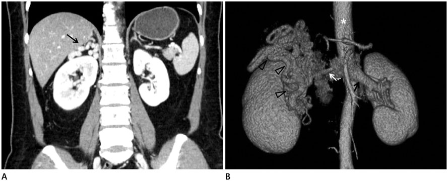
Fig. 1 Coronal reformatted CT image (A) and volume rendered image (B) of the aberrent right renal venous drainage. A. Contrast-enhanced coronal reformatted abdominal CT showed tortuous dilated vascular structure (black arrow) in the right suprarenal space and a normal parenchymal enhancement of both kidneys. B. Anterior volume-rendered image from CT data showed tortuous dilated vascular structure (open arrowheads) in the right renal hilum and suprarenal space. Whereas normal left renal vein (black arrow) is seen at hilar portion, the orthotopic right renal vein is not seen. The aorta (*) is seen and the right renal artery (white arrow) is normally seen.
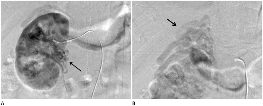
Fig. 2 Digital subtraction angiography (DSA) image showing the aberrent right renal venous drainage. A. In DSA image of the right renal arteriogram, the tortuous dilated vascular structure (black arrow) is visualized at hilar portion after the renal parenchyma was stained. The orthopedic renal vein is not observed at the expected location. B. In DSA image of the right renal arteriogram, the tortuous dilated vascular structure (black arrow) is visualized at the right suprarenal space just after the staining of hilar tortuous vessel. Still the orthopedic renal vein is not observed at the expected location.
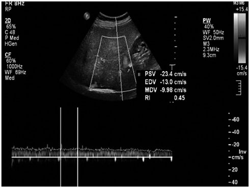
Fig. 3 Color and spectral Doppler ultrasound of vascular structure located at the right suprarenal space showed in the monophasic waveform, suggesting continuous venous flow.
Reference
-
1. Mathews R, Smith PA, Fishman EK, Marshall FF. Anomalies of the inferior vena cava and renal veins: embryologic and surgical considerations. Urology. 1999; 53:873–880.2. Satyapal KS. The renal veins: a review. Eur J Anat. 2003; 7:43–52.3. Bozlar U, Ugurel MS, Bedir S, Ors F, Coskun U, Aydur E. Right renal vein aplasia associated with diverted renal venous drainage through lower pole. Cardiovasc Intervent Radiol. 2008; 31:Suppl 2. S140–S143.4. Pinggera GM, Spranger R, Frauscher F, Eder R, Smekal A, Bartsch G. Congenital absence of the right renal vein. J Urol. 2003; 170:914–915.5. Satyapal KS. Classification of the drainage patterns of the renal veins. J Anat. 1995; 186(Pt 2):329–333.6. James EC, Fedde CW, Khuri NT, Gillespie JT. Division of the left renal vein: a safe surgical adjunct. Surgery. 1978; 83:151–154.7. Witz M, Korzets Z. Renal vein occlusion: diagnosis and treatment. Isr Med Assoc J. 2007; 9:402–405.8. Diniz GV, Pereira WJ, Moreira AC, Santos BM, Drumond DA, Petroianu A. Kidney function after left renal vein ligation in the dog. Rev Hosp Clin Fac Med Sao Paulo. 2001; 56:1–4.
- Full Text Links
-
- Actions
-
Cited
- CITED
-
- Close
- Share
-
- Similar articles
-
- Prevalence and clinical relevance of the anatomical variations of suprarenal arteries: a review
- Abnormal ramification pattern of the renal and testicular vessels
- Persisting subcardinal vein associated with unilateral ectopic pelvic kidney
- Absence of retromandibular vein associated with atypical formation of external jugular vein in the parotid region
- Levoatriocardinal Vein Combined with Pulmonary Venous Varix Mimicking Arteriovenous Malformations: A Case Report
