Anat Cell Biol.
2019 Dec;52(4):522-524. 10.5115/acb.19.071.
Persisting subcardinal vein associated with unilateral ectopic pelvic kidney
- Affiliations
-
- 1Department of Anatomy, Sri Lakshmi Narayana Institute of Medical Sciences, Puducherry, India. anat_rajesh@rediffmail.com
- KMID: 2466707
- DOI: http://doi.org/10.5115/acb.19.071
Abstract
- Absence of left kidney was noted during routine anatomy dissection of a male cadaver of South Indian origin. On examination of the abdomen and pelvic cavities; an ovoid mass of tissue was found in the pelvis, anterolateral to the sacrum. Further dissection revealed the presence of an ectopic left side kidney. The ectopic kidney was lying inferior to the sigmoid colon and anterior to the bifurcation of left common iliac vessel. It was supplied by numerous aberrant vessels from the terminal part of abdominal aorta. One of the renal veins which drain the ectopic kidney was found to be persisting subcardinal vein and it is a novel finding. Such ectopic pelvic kidneys are susceptible to blunt trauma, iatrogenic injuries as well as pathologic manifestations.
MeSH Terms
Figure
-
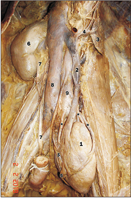
Fig. 1 Ectopic pelvic kidney with persisting subcardinal vein and aberrant renal arteries. 1, ectopic pelvic kidney; 2, persisting subcardinal vein; 3, suprarenal gland; 4, suprarenal vein; 5, pelvis of the ureter from ectopic kidney; 6, normal right kidney; 7, renal pelvis of normal kidney; 8, inferior vena cava; 9, abdominal aorta. Symbols * and $ indicate aberrant renal arteries and renal vein, respectively.
Reference
-
1. Standring S. Gray's anatomy: the anatomical basis of clinical practice. 41st ed. London: Elsevier Health Sciences;2016.2. Carlson BM. Human embryology and developmental biology. 4th ed. Philadelphia, PA: Mosby-Elsevier;2009.3. Chai OH, Song CH, Park SK, Kim W, Cho ES. Molecular regulation of kidney development. Anat Cell Biol. 2013; 46:19–31.4. Eid S, Iwanaga J, Loukas M, Oskouian RJ, Tubbs RS. Pelvic kidney: a review of the literature. Cureus. 2018; 10:e2775.5. Cinman NM, Okeke Z, Smith AD. Pelvic kidney: associated diseases and treatment. J Endourol. 2007; 21:836–842.6. Schmidlin FR, Iselin CE, Naimi A, Rohner S, Borst F, Farshad M, Niederer P, Graber P. The higher injury risk of abnormal kidneys in blunt renal trauma. Scand J Urol Nephrol. 1998; 32:388–392.7. Wadhwa P, Hemal AK. Case report: transmesocolic laparoscopic reconstruction of ureteropelvic junction obstruction in pelvic kidney associated with extrarenal calices. J Endourol. 2006; 20:188–190.8. Raghunath BV, Narendra BM, Gowrishankar BC, Ramesh S. Extrarenal calyces associated with pelviureteric junction obstruction: a case report of a rare anomaly. J Indian Assoc Pediatr Surg. 2012; 17:124–125.9. Gokalp G, Hakyemez B, Erdogan C. Vascular anomaly in bilateral ectopic kidney: a case report. Cases J. 2010; 3:5.10. Sadler TW. Langman's medical embryology. 14th ed. Philadelphia, PA: Wolters Kluwer;2019. p. 256–266.
- Full Text Links
-
- Actions
-
Cited
- CITED
-
- Close
- Share
-
- Similar articles
-
- A Case of Hypoplastic Ectopic Kidney in Pelvic Cavity
- A Case Report of Simple Unilateral Ectopic Kidney
- A Case of Unilateral Pelvic Kidney contained a Calculus
- Simple Unilateral Ectopic Kidney
- Four Cases of Unilateral Single Ectopic Ureter with Ipsilateral Dysplastic or Hypoplastic Kidney in Children: the Diagnostic Value of MR Urography
