Classic-Type Epithelioid Sarcoma Mimicking Multiple Droplet-Like Abscesses in the Lower Leg
- Affiliations
-
- 1Department of Radiology, Research Institute of Radiological Science, Medical Convergence Research Institute, and Severance Biomedical Science Institute, Yonsei University College of Medicine, Seoul, Korea. jss@yuhs.ac
- 2Department of Pathology, Yonsei University College of Medicine, Seoul, Korea.
- KMID: 2098020
- DOI: http://doi.org/10.3348/jksr.2015.72.6.405
Abstract
- Epithelioid sarcoma is an uncommon, slow-growing soft tissue tumor that usually arises in the distal part of an upper extremity, predominantly occurring in young adults. Classic-type epithelioid sarcomas appear to be less aggressive and have a better prognosis. In magnetic resonance imagng, epithelioid sarcoma usually appears as a subcutaneous mass or ulcer with cutaneous erosion. Here, we report a classic-type epithelioid sarcoma in a 68-year-old male without an apparent mass, presenting with only numerous small disseminated droplet-like ring-enhancing nodules mimicking abscesses, and multiple metastases.
MeSH Terms
Figure
-
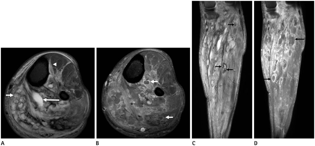
Fig. 1 MRI of the left lower leg. A. Axial T2-weighted fat saturation (TR/TE = 2468/70 ms) image shows multiple droplet-like hyperintense signal nodular lesion, mainly at superficial and deep posterior compartment of lower leg (short arrow). Diffuse muscular signal change was combined with focal fluid collection posterior to tibialis posterior (long arrow). The muscular signal change and nodular lesion was also noted at anterior compartment of leg (arrowhead). The margins of nodules were thin and had relatively regular thickness. B. Majority of these nodular lesions in axial gadolinium-enhanced T1-weighted fat saturation image (TR/TE = 618/20 ms) at the same section shows only peripheral ring-like enhancement (arrows). C. On coronal T2-weighted fat saturation (TR/TE = 2158/70 ms) image, there are disseminated nodular lesions from the level of proximal tibia to the level of distal tibia. Only in few, but some of nodules show internal intermediate or low signal foci (arrows). D. Coronal gadolinium-enhanced T1-weighted fat saturation (TR/TE = 618/20 ms) image at the same section shows numerous peripheral ring-like enhancing disseminated nodular lesions (arrows). TE = echo time, TR = repetition time
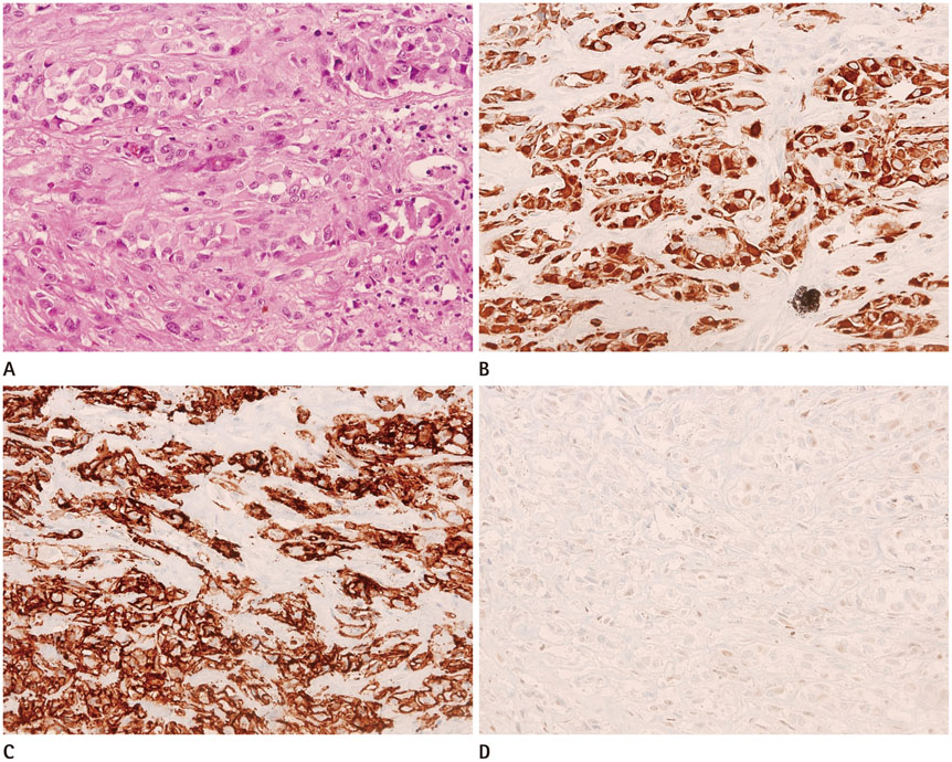
Fig. 2 Photomicrographies of histologic specimen. A. There are round to polygonal shape epithelioid cells with abundant eosinophilic cytoplasm and atypical nuclei possessing vesicular chromatin and small nucleoli (hematoxylin and eosin, × 200). B. Diffuse expression of cytokeratin are noted in the sarcoma (× 200). C. Strong membranous immunoreactivity for CD34 are noted (× 200). D. Complete loss of INI1 (SMARCB 1) protein expression in the tumor cells are noted (× 200).
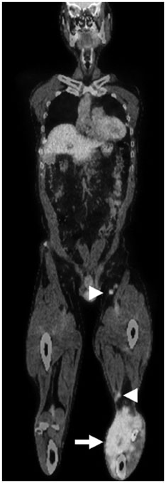
Fig. 3 On coronal fusion positron emission tomography/CT image, peripheral enhancing lesions of lower leg demonstrate markedly increased glucose metabolism (arrow). Ipsilateral popliteal and inguinal lymph nodes also show intense fluorodeoxyglucose uptake (maximum standardized uptake value, 6.85) (arrowheads).

Fig. 4 Chest CT scan shows numerous small nodules with random distribution in both lung parenchyme.
Reference
-
1. Hurtado RM, McCarthy E, Frassica F, Holt PA. Intraarticular epithelioid sarcoma. Skeletal Radiol. 1998; 27:453–456.2. Romero JA, Kim EE, Moral IS. MR characteristics of epithelioid sarcoma. J Comput Assist Tomogr. 1994; 18:929–931.3. Yamato M, Nishimura G, Yamaguchi T, Tamai K, Saotome K. Epithelioid sarcoma with unusual radiological findings. Skeletal Radiol. 1997; 26:606–610.4. von Hochstetter AR, Cserhati MD. Epithelioid sarcoma presenting as chronic synovitis and mistaken for osteosarcoma. Skeletal Radiol. 1995; 24:636–638.5. Dion E, Forest M, Brasseur JL, Amoura Z, Grenier P. Epithelioid sarcoma mimicking abscess: review of the MRI appearances. Skeletal Radiol. 2001; 30:173–177.6. Nakashima H, Katagiri H, Sugiura H, Yonekawa M, Nishida Y, Yamada Y. Epithelioid sarcoma mimicking a primary osseous multifocal scapula lesion. Skeletal Radiol. 2002; 31:430–433.7. Fletcher CDM. World Health Organization. International Agency for Research on Cancer. WHO classification of tumours of soft tissue and bone. 4th ed. Lyon: IARC Press;2013. p. 216–218.8. Rekhi B, Gorad BD, Chinoy RF. Clinicopathological features with outcomes of a series of conventional and proximal-type epithelioid sarcomas, diagnosed over a period of 10 years at a tertiary cancer hospital in India. Virchows Arch. 2008; 453:141–153.9. Tateishi U, Hasegawa T, Kusumoto M, Yokoyama R, Moriyama N. Radiologic manifestations of proximal-type epithelioid sarcoma of the soft tissues. AJR Am J Roentgenol. 2002; 179:973–977.10. Chao KC, Chen C, Hsieh SC, Fang CL, Lao WT, Chan WP. MRI of epithelioid sarcoma of the thigh. Clin Imaging. 2005; 29:60–63.
