J Korean Soc Radiol.
2012 Aug;67(2):113-116. 10.3348/jksr.2012.67.2.113.
Unexpected Metastasis of Intraductal Papillary Mucinous Neoplasm of the Bile Duct into Thoracic Cavity with Direct Extension: Case Report
- Affiliations
-
- 1Department of Radiology, Hanyang University Guri Hospital, Guri, Korea. radjena@hanyang.ac.kr
- 2Department of Radiology, Hanyang University Seoul Hospital, Seoul, Korea.
- KMID: 2097960
- DOI: http://doi.org/10.3348/jksr.2012.67.2.113
Abstract
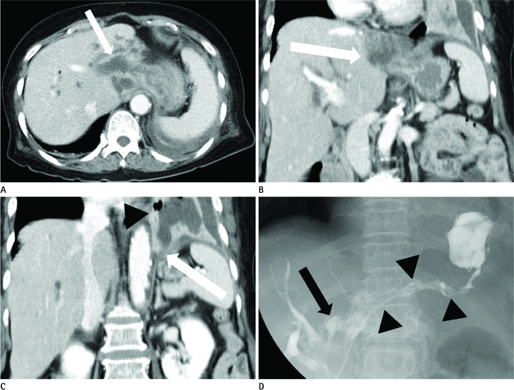
- Intraductal papillary mucinous neoplasm (IPMN) is known to arise from intraductal proliferation of mucinous cells with findings of marked dilatation of the biliary or pancreatic duct. There are reports of the metastasis and extension of pancreatic IPMN. However, cases of biliary IPMN with direct metastasis, or metastasis to distant locations, are rare. We present a case of metastasis of biliary IPMN with unexpected direct extension into the thoracic cavity, and we attempt to account for the mechanism of this extension.
Figure
Reference
-
1. Kloek JJ, van der Gaag NA, Erdogan D, Rauws EA, Busch OR, Gouma DJ, et al. A comparative study of intraductal papillary neoplasia of the biliary tract and pancreas. Hum Pathol. 2011; 42:824–832.2. Lim JH, Jang KT, Choi D. Biliary intraductal papillary-mucinous neoplasm manifesting only as dilatation of the hepatic lobar or segmental bile ducts: imaging features in six patients. AJR Am J Roentgenol. 2008; 191:778–782.3. Lim JH, Yoon KH, Kim SH, Kim HY, Lim HK, Song SY, et al. Intraductal papillary mucinous tumor of the bile ducts. Radiographics. 2004; 24:53–66. discussion 66-67.4. Kim HJ, Kim MH, Lee SK, Yoo KS, Park ET, Lim BC, et al. Mucin-hypersecreting bile duct tumor characterized by a striking homology with an intraductal papillary mucinous tumor (IPMT) of the pancreas. Endoscopy. 2000; 32:389–393.5. Yeh TS, Tseng JH, Chiu CT, Liu NJ, Chen TC, Jan YY, et al. Cholangiographic spectrum of intraductal papillary mucinous neoplasm of the bile ducts. Ann Surg. 2006; 244:248–253.6. Sugiyama M, Atomi Y. Intraductal papillary mucinous tumors of the pancreas: imaging studies and treatment strategies. Ann Surg. 1998; 228:685–691.7. Jang KT, Kwon GY, Ahn G. Clinicopathological analysis of growth patterns of malignant intraductal papillary mucinous tumors of the pancreas: unusual growth pattern of fistulous extension. Korean J Pathol. 2007; 41:38–43.8. Shibahara H, Tamada S, Goto M, Oda K, Nagino M, Nagasaka T, et al. Pathologic features of mucin-producing bile duct tumors: two histopathologic categories as counterparts of pancreatic intraductal papillary-mucinous neoplasms. Am J Surg Pathol. 2004; 28:327–338.9. Rosai J. Rosai and Ackerman's surgical pathology. 2004. Philadelphia: Elsevier;p. 1078–1080.10. Kaye MD. Pleuropulmonary complications of pancreatitis. Thorax. 1968; 23:297–306.
- Full Text Links
- Actions
-
Cited
- CITED
-
- Close
- Share
- Similar articles
-
- Intrahepatic and extrahepatic intraductal papillary neoplasms of bile duct
- Photodynamic Therapy Followed by Left Hepatectomy Used to Treat an Intraductal Papillary Mucinous Neoplasm of the Bile Duct
- Extrahepatic Bile Duct Duplication with Intraductal Papillary Neoplasm: A Case Report
- Intraductal Papillary Mucinous Tumor Simultaneously Involving the Liver and Pancreas: A Case Report
- Intraductal Papillary Mucinous Neoplasm of the Bile Ducts Accompanied by a Choledochoduodenal Fistula


