Amyloidoma of Retroperitoneal Lymph Nodes: A Case Report
- Affiliations
-
- 1Department of Radiology, College of Medicine, Hanyang University, Korea. songsy01@gmail.com
- 2Department of Radiology, College of Medicine, Hanyang University Guri Hospital, Korea.
- 3Department of Pathology, Hanyang University Hospital, College of Medicine, Korea.
- KMID: 2097948
- DOI: http://doi.org/10.3348/jksr.2011.64.3.261
Abstract
- Herein we report a case of retroperitoneal amyloidoma in a 56-year-old man and to describe its imaging findings and pathologic features. Abdomen computed tomography showed multiple nodular masses with amorphous calcifications in the retroperitoneum. On histologic review, these masses were composed of extensive nodular deposition of irregularly shaped amorphous eosinophilic material that was strongly positive on Congo red staining and apple green birefringence under polarizing microscopy, which is diagnostic for amyloidosis.
MeSH Terms
Figure
-
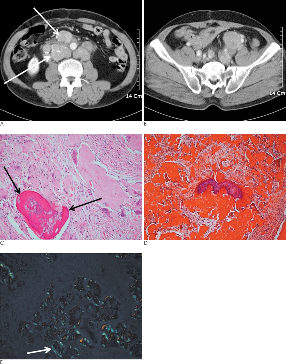
Fig. 1 A. An axial CT scan obtained at the level of the inferior pole of the right kidney. There are multiple nodular masses (see white arrows) around retroperitoneal great vessels with amorphous calcifications and surrounding infiltrations of low attenuation. B. At the level of the pelvic cavity, on axial CT image, an inhomogeneous enhancement pattern of amyloidoma is noted. C. Microscopic findings revealed a well demarcated mass that was composed of nodular depositions of irregularly shaped amorphous pale eosinophilic materials (see black arrows) and foci of calcification (H & E, ×200). These amyloid deposits were surrounded by multinucleated giant cells, histiocytes, lymphocytes, plasma cells, and ossification in dense fibrous stroma. D. Amyloid deposits were strongly positive on Congo red staining (×200). E. There is 'apple green' birefringence (white arrow) under polarizing microscopy (×200).
Reference
-
1. Sahoo S, Reeves W, DeMay RM. Amyloid tumor: a clinical and cytomorphologic study. Diagn Cytopathol. 2003; 28:325–328.2. Takebayashi S, Ono Y, Sakai F, Tamura S, Unayama S. Computed tomography of amyloidosis involving retroperitoneal lymph nodes mimicking lymphoma. J Comput Assist Tomogr. 1984; 8(5):1025–1027.3. Glynn TP Jr, Kreipke DL, Irons JM. Amyloidosis: diffuse involvement of the retroperitoneum. Radiology. 1989; 170:726.4. Borge MA, Parker LA, Mauro MA. Amyloidosis: CT appearance of calcified, enlarged periaortic lymph nodes. J Comput Assist Tomogr. 1991; 15:855–857.5. Krishnan J, Chu WS, Elrod JP, Frizzera G. Tumoral presentation of amyloidosis (amyloidomas) in soft tissues: a report of 14 cases. Am J Clin Pathol. 1993; 100:135–144.6. Georgiades CS, Neyman EG, Fishman EK. Cross-sectional imaging of amyloidosis: an organ system-based approach. J Comput Assist Tomogr. 2002; 26:1035–1041.7. Mohanty SK, Arora R, Kakkar N, Varma N, Panda N. Amyloidoma of lymph node. Am J of Hematol. 2002; 70:177–179.8. Georgiades CS, Neyman EG, Barish MA, Fishman EK. Amyloidosis: review and CT manifestations. Radiographics. 2004; 24:405–416.9. Ha CS, Medeiros LJ, Charnsangavej C, Crump M, Gospodarowicz MK. Lymphoma. Radiographics. 2006; 26:607–620.10. Pickhardt PJ, Bhalla S. Unusual nonneoplastic peritoneal and subperitoneal conditions: CT findings. Radiographics. 2005; 25:719–730.
- Full Text Links
-
- Actions
-
Cited
- CITED
-
- Close
- Share
-
- Similar articles
-
- Soft Tissue Amyloidoma of Upper Extremity: A Case Report
- A Case of Retroperitoneal Castleman's Disease
- A Case of Bilateral Trigeminal Amyloidoma Diagnosed Through an Endoscopic Transsphenoidal Approach
- A case of malignant lymphoma of testis, adrenal gland and retroperitoneal lymph nodes
- A case report of castleman disease in th neck: CT and MRI finding
