MR Imaging Features of Primary Cutaneous Diffuse Large B-cell Lymphoma: A Case Report
- Affiliations
-
- 1Department of Diagnostic Radiology, College of Medicine, Yeungnam University, Korea. sungho1999@ynu.ac.kr
- KMID: 2097946
- DOI: http://doi.org/10.3348/jksr.2011.64.3.249
Abstract
- Here we report magnetic resonance (MR) imaging findings for a case of primary cutaneous diffuse large B-cell lymphoma (PCDLBCL) located in the chest wall, a rare type of lymphoma, occurring in a 67-year-old woman. Although rare, PCDLBCL may be one of the diagnostic inclusions for a soft tissue mass in the subcutaneous layer of the trunk, especially when the mass shows hyperintensity in relation to the muscle on T2-weighted MR images and peripheral enhancement on contrast enhanced T1-weighted MR images.
MeSH Terms
Figure
-
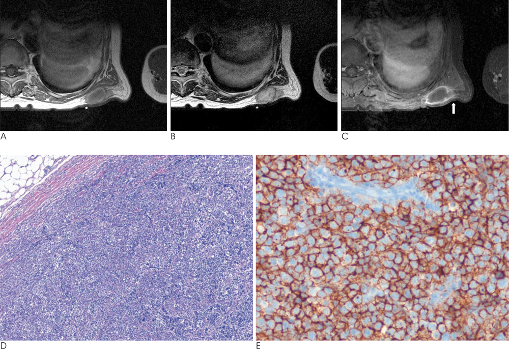
Fig. 1 67-year-old woman with painful primary cutaneous diffuse large B-cell lymphoma in the left posterior chest wall. A. This Axial T1-weighted image shows a hypointense lesion and a peripheral high signal intensity rim on the chest wall. B. An axial T2-weighted image showing a lobulating subcutaneous mass with a central high signal intensity and peripheral low signal intensity rim. C. A gadolinium-enhanced, fat-suppressed, T1-weighted image that shows a mass with thick and irregular peripheral enhancement. Subtle subcutaneous and overlying skin enhancements (arrow) are present near the mass. D. Photomicrography of the skin shows diffuse dermal infiltration by large atypical lymphocytes (H & E stain, ×40). E. Immunohistochemical staining of tumor cells showing expression of CD20.
Reference
-
1. Narimatsu H, Morishita Y, Shimada K, Ozeki K, Kohno A, Kato Y, et al. Primary cutaneous diffuse large B cell lymphoma: a clinically aggressive case. Intern Med. 2003; 42:354–357.2. Hwang S. Imaging of lymphoma of the musculoskeletal system. Radiol Clin North Am. 2008; 46:379–396.3. Goodlad JR, Krajewski AS, Batstone PJ, Mckay P, White JM, Benton EC, et al. Primary cutaneous diffuse large B-cell lymphoma: prognostic significance of clinicopathological subtypes. Am J Surg Pathol. 2003; 27:1538–1545.4. Hallermann C, Niermann C, Fischer RJ, Schulze HJ. New prognostic relevant factors in primary cutaneous diffuse large B-cell lymphomas. J Am Acad Dermatol. 2007; 56:588–597.5. Chen YF, Li YC, Chen LM, Tu CC, Chang CC, Kuo SY, et al. Primary cutaneous diffuse large B cell lymphoma relapsed solely as a huge lung tumor mimicking a primary pulmonary lymphoma. Int J Hematol. 2010; 91:112–116.6. Sharon V, Mecca PS, Steinherz PG, Trippett TM, Myskowski PL. Two pediatric cases of primary cutaneous B-cell lymphoma and review of the literature. Pediatr Dermatol. 2009; 26:34–39.7. Willemze R, Jaffe ES, Burg G, Cerroni L, Berti E, Swerdlow SH, et al. WHO-EORTC classification for cutaneous lymphomas. Blood. 2005; 105:3768–3785.8. Hembury TA, Lee B, Gascoyne RD, Macpherson N, Yang B, House N, et al. Primary cutaneous diffuse large B-cell lymphoma: a clinicopathologic study of 15 cases. Am J Clin Pathol. 2002; 117:574–580.9. ter Braak PM, Guit GL, Bloem JL. Case 111: Soft-tissue lymphoma. Radiology. 2007; 243:293–296.10. Hong SH, Chung HW, Choi JY, Koh YH, Choi JA, Kang HS. MRI findings of subcutaneous epidermal cysts: emphasis on the presence of rupture. AJR Am J Roentgenol. 2006; 186:961–966.
- Full Text Links
-
- Actions
-
Cited
- CITED
-
- Close
- Share
-
- Similar articles
-
- A Case of Primary Cutaneous Diffuse Large B-cell Lymphoma
- A Case of Primary Cutaneous Diffuse Large B-cell Lymphoma
- Primary Cutaneous T-cell/histiocyte-rich B-cell Lymphoma
- A Case of Secondary Cutaneous Diffuse Large B-cell Lymphoma
- A Case of Primary Cutaneous Diffuse Large B-cell Lymphoma,Leg Type Which Metastasized to the Brain
