Correlation among Magnetic Resonance Images, Electron Microscopic Findings, Light Microscopic Findings and Clinical Symptom of the Degeneration of Lumbar Intervertebral Disc
- Affiliations
-
- 1Department of Orthopedic Surgery, School of Medicine, Chungnam National University, Taejon, Korea. jsahn@cuvic.cnu.ac.kr
- 2Department of Pathology, School of Medicine, Chungnam National University, Taejon, Korea.
- KMID: 2097751
- DOI: http://doi.org/10.4184/jkss.2001.8.2.121
Abstract
-
STUDY DESIGN: A retrospective study was performed in patients who had undergone any operation with removal of lumbar intervertebral disc at Chungnam National University Hospital.
OBJECTIVES
To evaluate relationship among magnetic resonance image, electron microscopic findings, light microscopic findings and clinical symptoms in degenerated intervertebral disc. SUMMARY OF LITERATURE REVIEW: Degenerative changes and disc herniations in the intervertebral disc have been shown to be accompanied by changes in the water and proteoglycan content of the tissue.
MATERIALS AND METHODS
Our study followed by any operation with removal of intervertebral disc was carried out on 60 patients at Chungnam National University Hospital from January 1998 to December 1999. In radiographic evaluation we used a criteria from Frymoyer. In clinical evaluation we classified clinical symptom according to scale of Kirkaldy-Willis. And we classified of electron microscopic findings into five grades according to degrees of denudation of proteoglycan from hyalunonic acid. In light microscopic findings, we classified by cell nest formation, noevascularization and amount of muccopolysaccharide.
RESULTS
In radiologic evaluation there were 11 cases in grade III, 28 cases in grade IV, and 21 cases in grade V. There were no grade I, II in our study. In clinical symptom, there were 20 cases in Good, 18 cases in Fair, and 22 cases in Poor. In electron micro-scopic findings, there were 4 cases in grade 3, 35 cases in grade 4, and 21 cases in grade 5. There were no grade I, II in this study. There was a relationship between magnetic resonance image and electron microscopic findings and clinical symptom (p<0.05).
CONCLUSION
Our study of electron microscopic findings of degenerated intervertebral disc may be a help to understand of pathogenesis of disc prolapse.
MeSH Terms
Figure
-
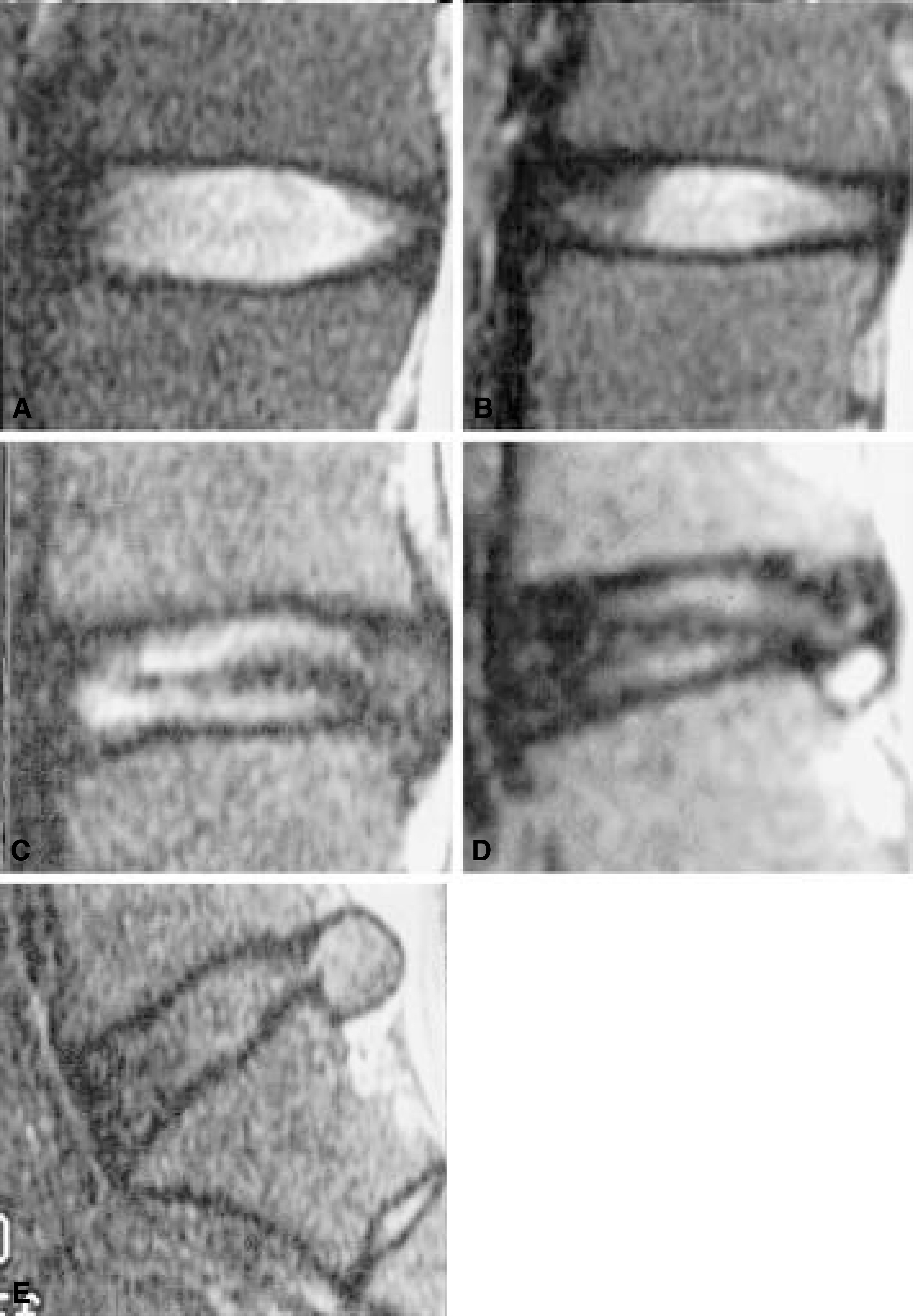
Fig. 1. MRI findings showing pattern of each grade of degeneration of nucleus pulposus Fig. A. Grade I-Homogenous bright Fig. B. Grade II-Horizontal dark band Fig. C. Grade III-Gray tone with dark stippling Fig. D. Grade IV-Almost dark tone with bright stippling Fig. E. Grade V-Total darkness or gross loss of disc height

Fig. 2. EM finding showing Grade III(From 41 to 60% of proteoglycan are denuded from hyarulonic acid).
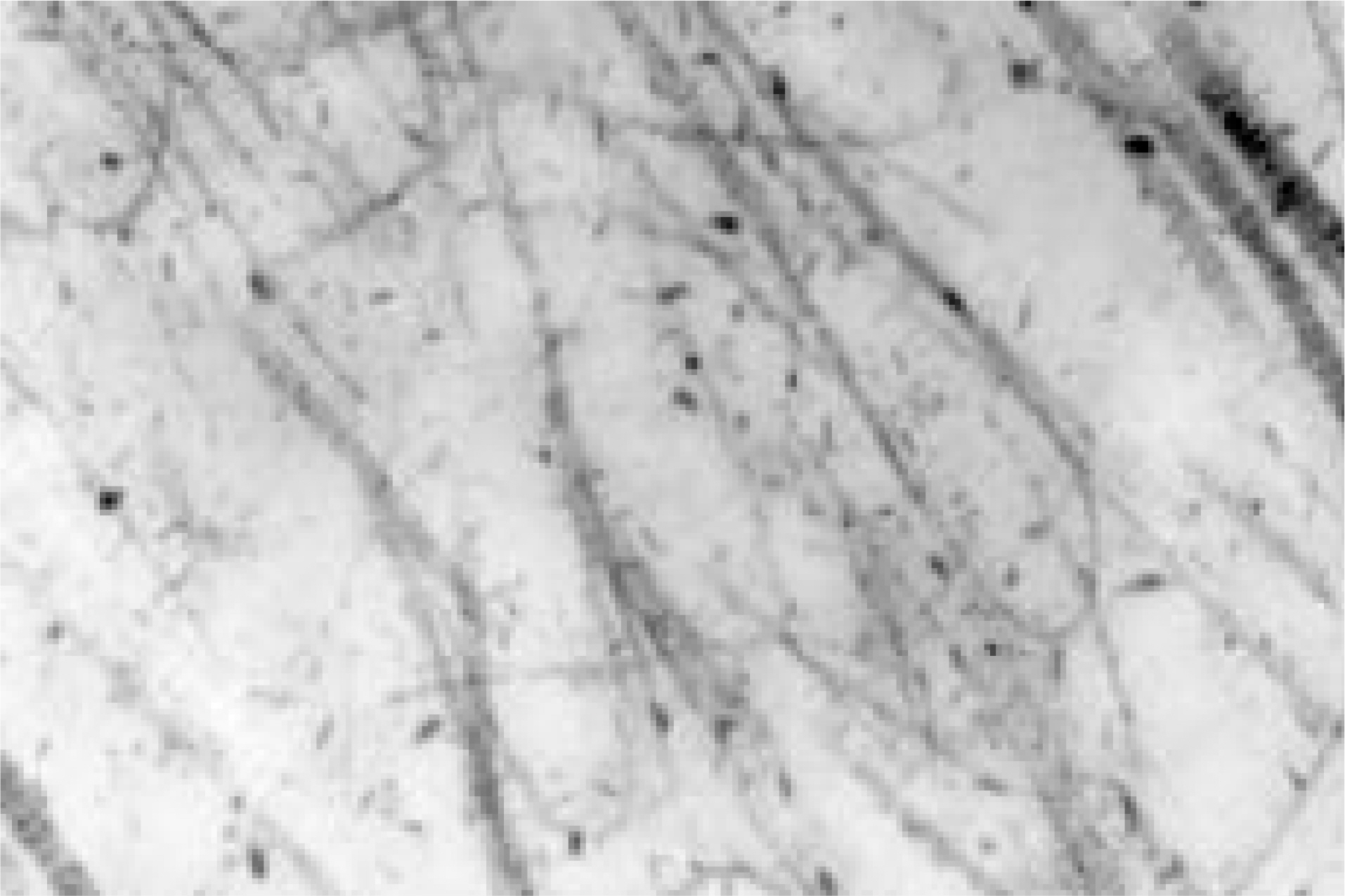
Fig. 3. EM finding showing Grade IV(From 61 to 80% of proteoglycan are denuded from hyarulonic acid).
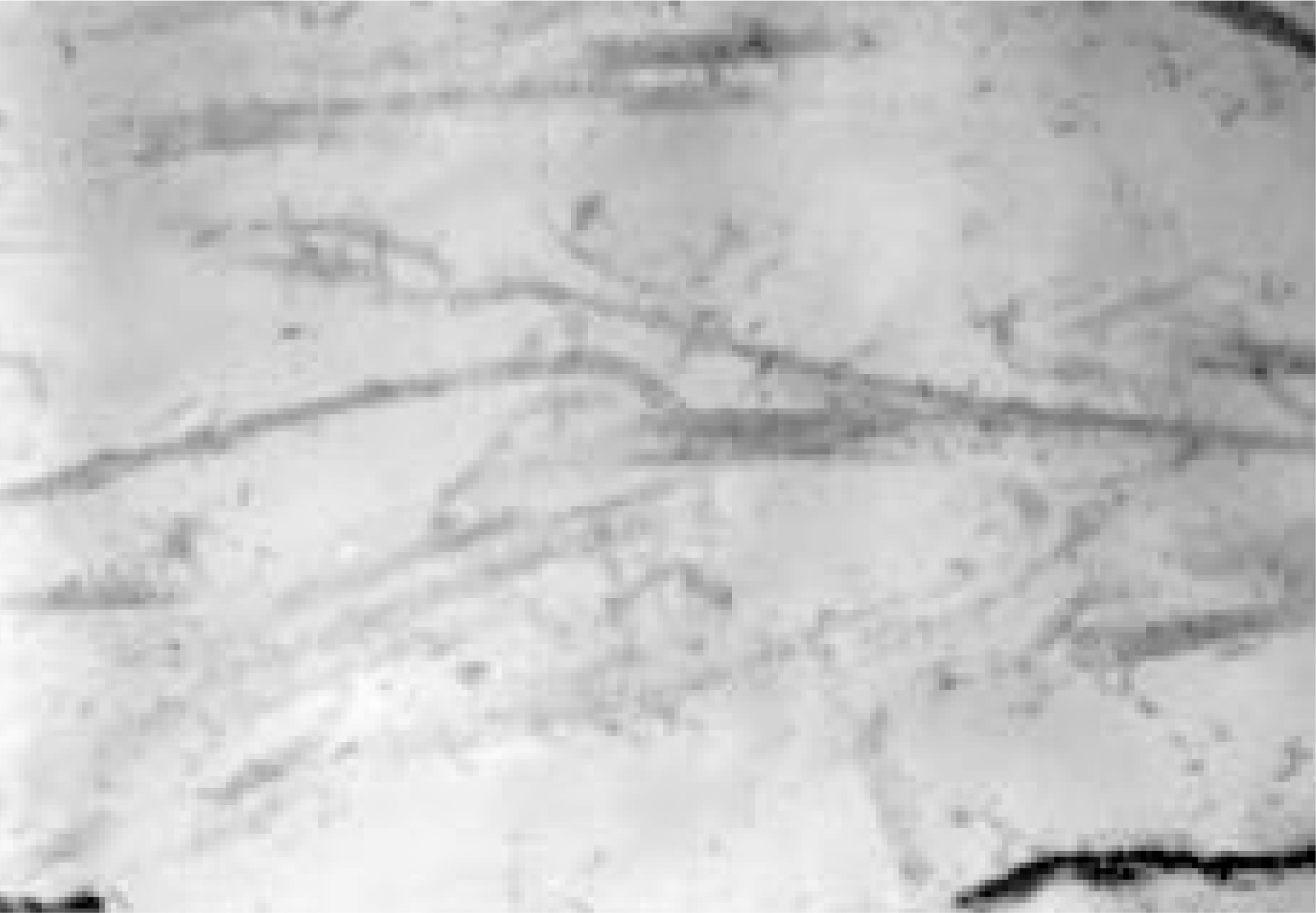
Fig. 4. EM finding showing Grade V(Over 81% of proteoglycan are denuded from hyarulonic acid).
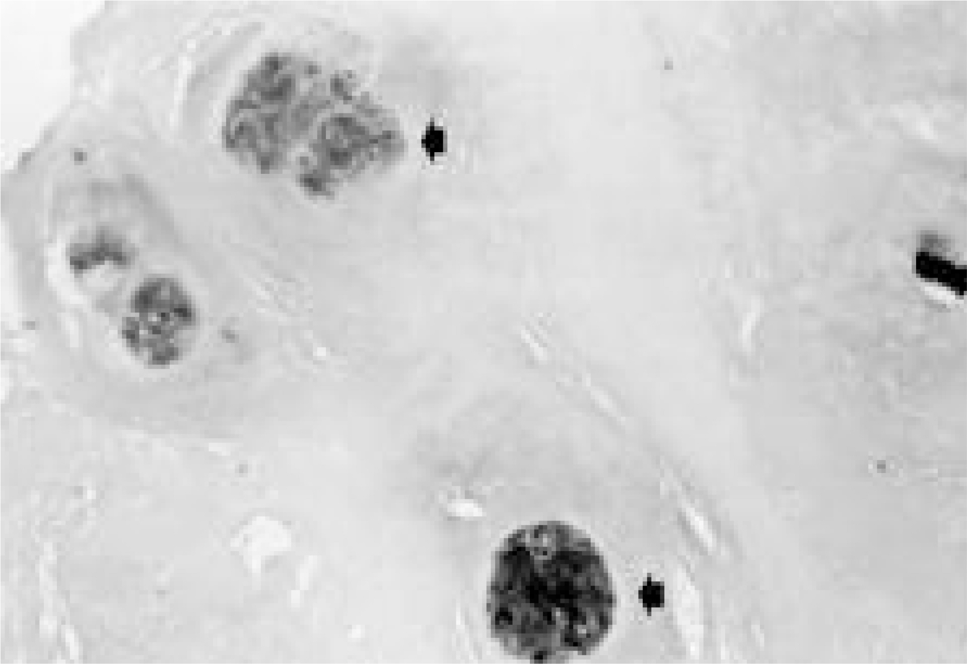
Fig. 5. LM finding: Disc tissue showing cell nest formation (HE stain, × 400).
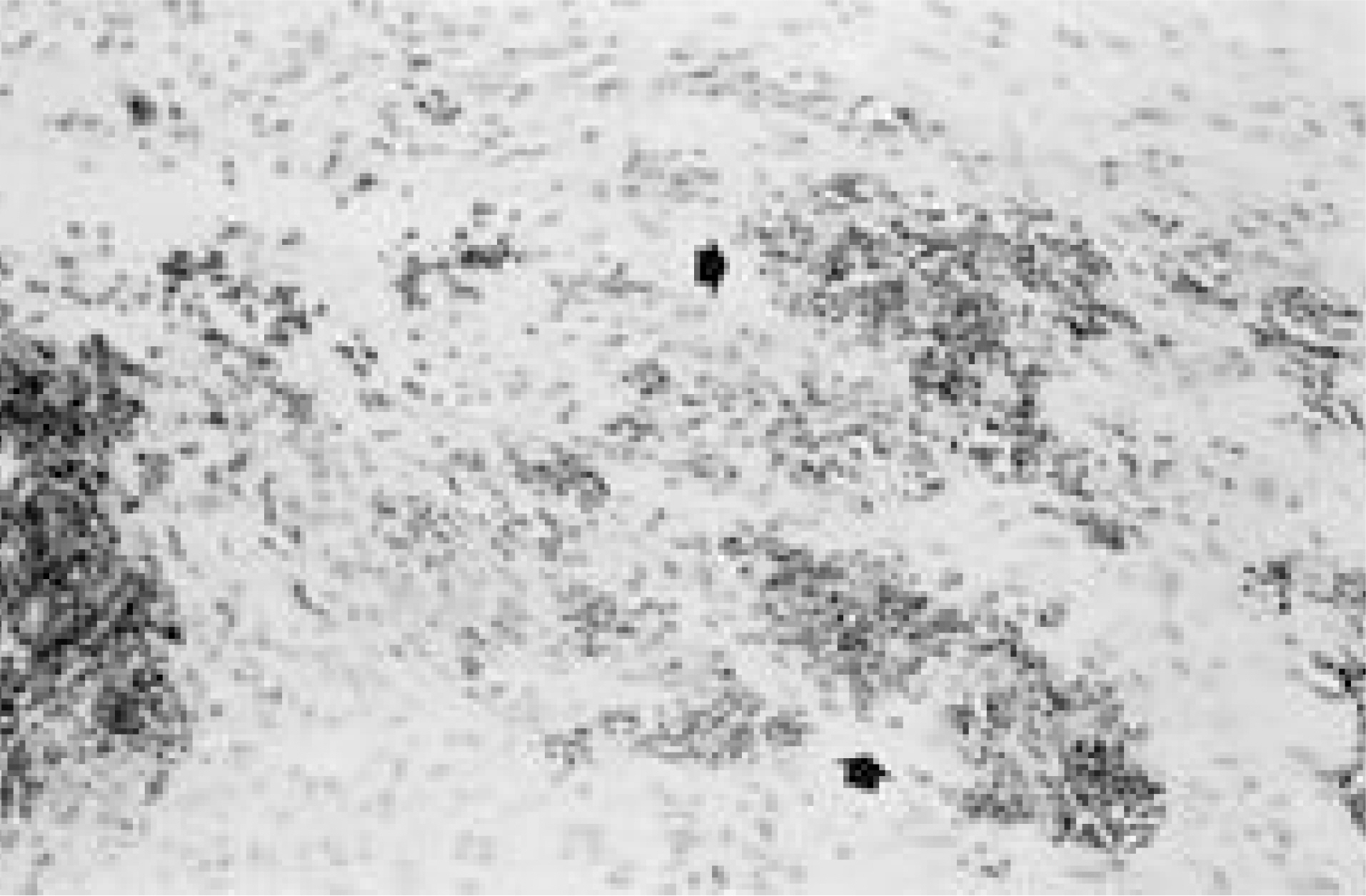
Fig. 6. LM finding: Disc tissue showing blood vessels (noevascularization) (HE stain, × 200).
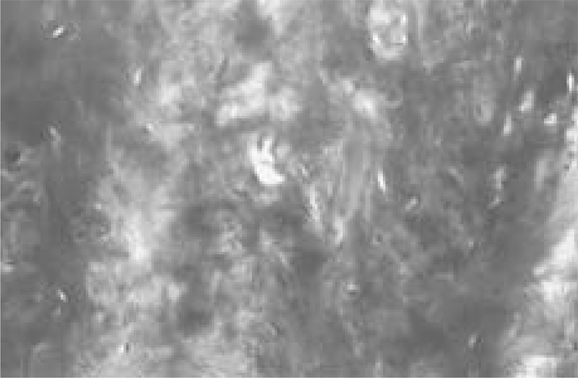
Fig. 7. LM finding: Disc tissue shows strong positive for neutral muccopolysaccharide in PAS stain (PSA stain, × 200).
Reference
-
1). Adams P, Muir H. Qualitive changes with age of proteoglycans of human lumbar disc. Ann Rheum Dis. 35:289–296. 1976.2). Bernick S, Walker JM, Paule WJ. A ge changes to the anulus fibrosis in human intervertebral disc, Spine. 16:520–524. 1991.3). Bishop PB, Pearce RH. The proteoglycans of the carti -laginous endplate of the human intervertebral disc change after maturity. J Orthop Res. 11:324–331. 1993.4). Brock M, Patt A, Mayer HM. The form and structure of the extruded disc. Spine. 17:1547–1461. 1992.
Article5). Buckwalter JA. Aging and degeneration of the human intervertebral disc. Spine. 20:1307–1314. 1995.
Article6). Buckwalter JA. The fine structure of the human intervertebral disc. In: Symposium on idiopathic low back pain. ed by AA White, SL Gorden, St Louis, CV Mosby Co.:. 108–143. 1982.7). Buckwalter JA, Pedrini-Mille A, Pedrini V, et al. .:. Proteoglycans of human infant intervertebral disc-Electron microscopic and Biochemical studies. J Bone Joint Surg. 67(A):284–294. 1985.8). Buckwalter JA, Smith KC, Kazarien LE, et al. .:. Articular cartilage and intervertebral disc proteoglycans differ in structure: An electron microscopic study. J Orthop Res. 7:141–151. 1989.
Article9). Conventry MB, Ghormley RK, Kernohan JW. The intervertebral disc: Its microscopic anatomy and pathology. I. Anatomy, development, and physiology. J Bone Joint Surg. 27:105–112. 1945.10). Donohue PJ, Jahnke MR, Blaha MD, et al. Character -ization of link protein(s) from human intervertebral-disc tissue. Biochem J. 251:739–747. 1988.11). Eyre DR. Biochemistry of the intervertebral disc. Int Rev Conn Tiss Res. 8:227–291. 1979.
Article12). Eyre DR, Benya PD, Buckwalter JA, et al. Intervertebral disk-basic science perspectives, In: New Perspectives on Low Back Pain.ed by JW Frymoyer, SL Gorden, Park Ridge, IL: AAOS:147-195. 1989.13). Eyre DR, Muir H. Quantitative analysis of typee I and II collagens in human intervertebral discs at various ages. Biochim Biophys Acta. 492:29–42. 1977.14). Eyre DR, Muir H. Types I and II collagens in interverte bral disc: Interchanging radial distributions in annulus fibrosus. Biochem J. 157:267–270. 1976.15). Frymoyer JW. The adult spine-Principles and Practice-,2nd ed. Philadelphia: Lippincott-Raven Publisher;p. 741–742. 1997.16). Galante JO. Tensile properities of the human lumbar annulus fibrosus. Acta Orthop Scand. 100(suppl):1–91. 1967.17). Gibson MJ, Buckley J, Mawhinney R, et al. .:. Magnetic resonance imaging and discography in diagnosis of disc degenerstion. J Bone Joint Surg. 68(B):369–373. 1986.18). Hardingham TE. The role of link protein in the structure of cartilage proteoglycan aggregates. Biochim J. 177:237–247. 1979.19). Ishii T, Tsuji H, Sano A, et al. .:. Histochemical and ultra -structural observations on brown degeneration of human intervertebral disc. J Orthop Res. 9:78–90. 1991.20). Jeon CH, Kim HK, Kang SY. The study for the neovas -cularization and basic fibroblast growth factor (bFGF) Expression in the intervertebral disc tissue associated with aging and disc degeneration. J of Korean spine surg. 6:329–335. 1999.21). Kang CN, Wang JM, Rho KJ, et al. .:. The relationships between the radiologic degenerative changes and histologic changes in herniated intervertebral disc. J of Korean Orthop Assoc. 27:1244–1255. 1992.22). Kim YT, Kim JJ, Hwang JH, et al. .:. Annulus tears in the intervertebral disc degeneration-An experimental study using an animal model-, J of Korean spine surg. 1:259–272. 1994.23). Kr mer J. Intervertebral disk diseases. Causes, diagnosis, treatment and prophylaxis. 2nd ed.New York: Thieme medical publishers Inc;p. 41–51. 1990.24). Luoma K, Vehmas T, Riihimaki H, et al. Disc height and signal intensity of the nucleus pulposus on magnetic resonance imaging as indicators of lumbar disc degeneration. Spine. 26:680–686. 2001.
Article25). Luoma K, Riihimaki H, Luukkonen R, et al. Low back pain in relation to lumbar disc degeneration. Spine. 25:487–492. 2000.
Article26). Lyons G, Eisenstein SM, Sweet MB. Biochim Biophys Acta. 673:443–453. 1981.27). Maroudas A. Physical chemistry of articular cartilage and the intervertebral disc. The Joint and Synovial fluid. Vol. II:ed by. L Sokoloff, editor. Orlando: Academic Press;p. 239–291. 1980.
Article28). Mow VC, Zhu WB, Lai WM, et al. .:. The influence of link protein stabilization on the viscometric properties of proteoglycan aggregate solutions. Biochim Biophys Acta. 992:201–208. 1989.
Article29). Naylor A. The biochemical changes in the human intervertebral disc in degeneration and nuclear prolapse. Clin N Am. 2:343–358. 1971.
Article30). Olczyk K. Age-related changes in proteoglycans of human intervertebral discs. J Rheumatol. 53:19–25. 1994.31). Osti OL, Fraser RD. MRI and discography of annular tears and intervertebral disc degeneration. J Bone Joint Surg. 74(B):431–435. 1992.32). Pearce RH, Grimmer BJ. The chemical constitution of the proteoglycan of human intervertebral disc. Biochem J. 157:753–763. 1976.
Article33). Pearce RH, Grimmer BJ, Adams ME. De generat ed and the chemical composition of the human lumbar intervertebral disc. J Orthop Res. 5:198–205. 1987.34). Pearce RH, Mathieson JM, Mort JS, et al. .:. Effect of age on the abundance and fragmentation of link protein of the human intervertebral disc. J Orthop Res. 7:861–867. 1989.
Article35). Pearce RH, Thompson JP, Bebault GM, et al. .:. Magnetic resonance imaging reflects the chemical changes of aging degeneration in the human intervertebral disk. J Rheumatol suppl. 27:42–43. 1991.36). Pearson CH, Happey F, Nayloe A, et al. .:. Collagens and associated glycopreteins in the human intervertebral disc. Ann Rheum Dis. 31:45–53. 1972.37). Pritzker KPH. Aging and degeneration in the lumbar intervertebral disc. Orthop Clin N Am. 8:65–77. 1977.
Article38). Schiebler ML, Camerino VJ, Fallon MD, et al. .:. In vivo and ex vivo magnetic resonance imaging evaluation of early disc degeneration with histopathologic correlation. Spine. 16:635–640. 1991.
Article39). Schneiderman G, Flannigan B, Kingston S, et al. .:. Magnetic resonance imaging in the diagnosis of disc degeneration: correlation with discography. Spine. 12:276–281. 1987.40). Southern EP, Fye MA, Panjabi MM, et al. .:. Disc degeneration: a human cadaveric study correlating magnetic resonance imaging and quantitative discomanometry. Spine. 25:2171–2175. 2000.41). Stevens RL, Dondi PG, Muir H. Proteoglycans of the intervertebral disc. Biochem J. 179:573–578. 1979.42). Tengblad A, Pearce RH, Grimmer BJ. Demonstration of link protein in protein aggregates from human intervertebral disc. Biochem J. 222:85–92. 1984.43). Urban JPG, Holm SH. I ntervertebral disc nutrition as related to spinbal movement and fusion, In: Tissue Nutrition and Viability. AR Hargens, editor. New York: Springer-Ver;ag;1985.44). Urban JPG, Holm SH, Marouds A, et al. .:. Nutrition of the IVD - as in vivo study of solute transport. Clin Orthop Rel Res. 129:101–141. 1977.45). Urban JPG, Maroudas A. The chemistry of the intervertebral disc in relation to its physiological function and requirements. Clin Rheum Dis. 6:51–76. 1980.
Article46). Wang JM, Kim DJ. Diagnostic value of image findings of MRI and discography for the internal disc disruption. J of Korean Orthop Assoc. 32:497–505. 1997.
Article47). Wang JM, Kim DJ. Diagnostic value of image findings of MRI for the intervertebral disc disruption, J of Korean Spine Surg. 4:36–42. 1997.48). Weidenbaum M, Foster RJ, Best BA, et al. .:. Correlating magnetic imaging with the biochemical content of the normal human intervertebral disc. J of Orthop Res. 10:552–561. 1992.49). Weidner N, Rice DT. Intervertebral disk material criteria for determining probable prolapse. Human Pathology. 19:406–410. 1988.
Article
- Full Text Links
-
- Actions
-
Cited
- CITED
-
- Close
- Share
-
- Similar articles
-
- Correlation of Magnetic Resonance Imaging of Lumbar Herniated Intervertebral Disc with Operative Findings
- The Classification of Lumbar Interspinous Ligament in Relation to Herniated Intervertebral Disc and Spinal Degeneration of Korean
- Lumbar Disc Degeneration and Segmental Instability: A Comparison of Magnetic Resonance Images and Plain Radiographs
- Correlation between Bone Mineral Density and Interverterbral Disc Degeneration
- Spinal Nerve Root Swelling Mimicking Intervertebral Disc Herniation in Magnetic Resonance Imaging: A Case Report
