J Korean Surg Soc.
2011 Jun;80(Suppl 1):S43-S46. 10.4174/jkss.2011.80.Suppl1.S43.
A giant retroperitoneal lymphangioma in a patient with neurofibromatosis type 1
- Affiliations
-
- 1Department of Medicine, Eulji University Hospital, Eulji University College of Medicine, Daejeon, Korea. minogiya@dreamwiz.com
- 2Department of Surgery, Eulji University Hospital, Eulji University College of Medicine, Daejeon, Korea.
- 3Department of Radiology, Eulji University Hospital, Eulji University College of Medicine, Daejeon, Korea.
- 4Department of Pathology, Eulji University Hospital, Eulji University College of Medicine, Daejeon, Korea.
- KMID: 2096649
- DOI: http://doi.org/10.4174/jkss.2011.80.Suppl1.S43
Abstract
- Neurofibromatosis type 1 (NF-1) is a genetically inherited disorder that may cause skin abnormalities and tumors that form on nerve tissues. These tumors can be small or large and can occur anywhere in the body, including the brain, spinal cord, or other peripheral nerves. Retroperitoneal lymphangiomas are very rare benign malformations of the lymphatic system. About 95% lymphangiomas occur in the skin and the subcutaneous tissues of the head, neck and axillary region and the remaining 5% appear in other parts of the body such as lungs, pleura, pericardium, liver, gallbladder, kidney, and the mesentery. Herein, we report the case of a giant retroperitoneal lymphangioma in a patient with NF-1 with a review of the literature.
MeSH Terms
Figure
-

Fig. 1 (A) Chest computed tomography (CT) shows multifocal soft tissue masses with calcification in the bilateral axillae, right pectoralis muscle, intermuscular layer of the left posterolateral chest wall. (B) Abdominal CT findings. It shows retroperitoneal giant cystic mass.
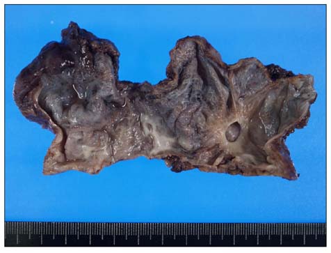
Fig. 2 Gross findings shows lymphangioma.
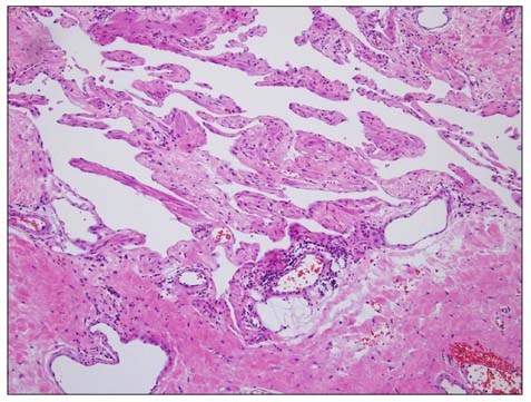
Fig. 3 Microscopic findings show lymphangioma. Proliferation of anastomosing blood vessels and smooth muscle (H&E, ×400).
Reference
-
1. Kurtz RJ, Heimann TM, Holt J, Beck AR. Mesenteric and retroperitoneal cysts. Ann Surg. 1986. 203:109–112.2. Takiff H, Calabria R, Yin L, Stabile BE. Mesenteric cysts and intra-abdominal cystic lymphangiomas. Arch Surg. 1985. 120:1266–1269.3. Torpy JM, Burke AE, Glass RM. JAMA patient page. Neurofibromatosis. JAMA. 2009. 302:2170.4. Lu-Emerson C, Plotkin SR. The neurofibromatoses. Part 1: NF1. Rev Neurol Dis. 2009. 6:E47–E53.5. Yang DM, Jung DH, Kim H, Kang JH, Kim SH, Kim JH, et al. Retroperitoneal cystic masses: CT, clinical, and pathologic findings and literature review. Radiographics. 2004. 24:1353–1365.6. Rieker RJ, Quentmeier A, Weiss C, Kretzschmar U, Amann K, Mechtersheimer G, et al. Cystic lymphangioma of the small-bowel mesentery: case report and a review of the literature. Pathol Oncol Res. 2000. 6:146–148.7. Allen JG, Riall TS, Cameron JL, Askin FB, Hruban RH, Campbell KA. Abdominal lymphangiomas in adults. J Gastrointest Surg. 2006. 10:746–751.8. Ordóñez NG. Value of immunohistochemistry in distinguishing peritoneal mesothelioma from serous carcinoma of the ovary and peritoneum: a review and update. Adv Anat Pathol. 2006. 13:16–25.9. Méndez-Gallart R, Solar-Boga A, Gómez-Tellado M, Somoza-Argibay I. Giant mesenteric cystic lymphangioma in an infant presenting with acute bowel obstruction. Can J Surg. 2009. 52:E42–E43.10. Jain R, Bandhu S, Sawhney S, Mittal R. Sonographically guided percutaneous sclerosis using 1% polidocanol in the treatment of vascular malformations. J Clin Ultrasound. 2002. 30:416–423.
