Piriformis Muscle: Clinical Anatomy with Computed Tomography in Korean Population
- Affiliations
-
- 1Department of Anesthesiology and Pain Medicine, Daegu Wooridul Hospital, Daegu, Korea.
- 2Department of Neurosurgery, Wooridul Hospital, Seoul, Korea.
- 3Department of Anesthesiology and Pain Medicine, Seoul National University Hospital, Seoul, Korea.
- 4Department of Anesthesiology and Pain Medicine, School of Medicine, Ewha Womans University, Seoul, Korea. ingoo97@lycos.co.kr
- KMID: 2074003
- DOI: http://doi.org/10.3344/kjp.2011.24.2.87
Abstract
- BACKGROUND
The objective was to evaluate the distance from the skin and the diameter of the piriformis muscle and their relationship to the body mass index (BMI).
METHODS
The study was a prospective study involving 60 patients. Patients were prepared on a radiological table in the prone position. Several images were obtained of each. In this view, the distance between the subcutaneous tissue and the piriformis muscle, and the diameter of the piriformis, were measured at three points (medially to laterally).
RESULTS
The distance to the piriformis from the skin was 6.6 +/- 0.9 cm, 6.3 +/- 0.8 cm, and 5.2 +/- 0.9 cm in terms of the lateral, center, and medial measurement, respectively. The center of the piriformis had a greater diameter with 1.7 +/- 0.4 (0.9-2.5) cm. The distance to the piriformis increased with BMI.
CONCLUSIONS
This study shows that the lateral of the piriformis muscle has a relatively greater distance from the skin. The center of the piriformis showed a greater diameter than other two portions. We found that the distance of the piriformis from subcutaneous tissues was correlated with BMI, but the diameter of the piriformis was not affected by BMI. These measurements can be used as a reference for determining the piriformis injection site in patients with piriformis syndrome.
Keyword
MeSH Terms
Figure
-

Fig. 1 This figure shows the measurement in this study. The (A), (B), and (C) represent the skin entry distance from medial, central, and lateral respectively. A right piriformis muscle seen in the computed tomography (arrow).
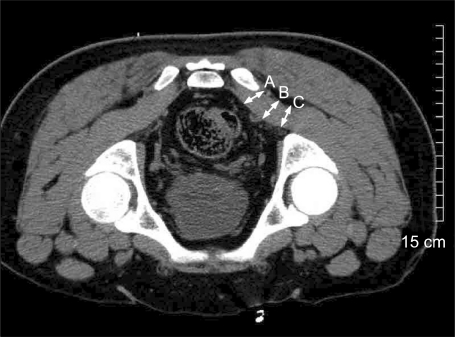
Fig. 2 This figure shows the measurement in this study. The (A), (B), and (C) represent the diameter of the piriformis from medial, center, and lateral respectively. Computed tomography views of right piriformis muscle (arrow).
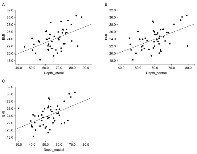
Fig. 3 Correlation between body mass index and distance to the piriformis. (A) Correlation between body mass index and distance to lateral portion, (B) correlation between body mass index and distance to central portion of the piriformis (r = 0.510, P = 0.000). (C). Correlation between body mass index and distance to medial portion of the piriformis (r = 0.481, P = 0.001).
Cited by 1 articles
-
Extra-spinal sciatica and sciatica mimics: a scoping review
Md Abu Bakar Siddiq, Danny Clegg, Suzon Al Hasan, Johannes J Rasker
Korean J Pain. 2020;33(4):305-317. doi: 10.3344/kjp.2020.33.4.305.
Reference
-
1. Ozaki S, Hamabe T, Muro T. Piriformis syndrome resulting from an anomalous relationship between the sciatic nerve and piriformis muscle. Orthopedics. 1999; 22:771–772. PMID: 10465490.
Article2. Hallin RP. Sciatic pain and the piriformis muscle. Postgrad Med. 1983; 74:69–72. PMID: 6878094.
Article3. Parziale JR, Hudgins TH, Fishman LM. The piriformis syndrome. Am J Orthop (Belle Mead NJ). 1996; 25:819–823. PMID: 9001677.4. Pace JB, Nagle D. Piriform syndrome. West J Med. 1976; 124:435–439. PMID: 132772.5. Chen WS. Bipartite piriformis muscle: an unusual cause of sciatic nerve entrapment. Pain. 1994; 58:269–272. PMID: 7816495.
Article6. Sayson SC, Ducey JP, Maybrey JB, Wesley RL, Vermilion D. Sciatic entrapment neuropathy associated with an anomalous piriformis muscle. Pain. 1994; 59:149–152. PMID: 7854796.
Article7. Jeon SY, Moon HS, Han YJ, Sung CH. Post-radiation piriformis syndrome in a cervical cancer patient: a case report. Korean J Pain. 2010; 23:88–91. PMID: 20552082.
Article8. Fishman LM, Zybert PA. Electrophysiologic evidence of piriformis syndrome. Arch Phys Med Rehabil. 1992; 73:359–364. PMID: 1554310.
Article9. Jankiewicz JJ, Hennrikus WL, Houkom JA. The appearance of the piriformis muscle syndrome in computed tomography and magnetic resonance imaging. A case report and review of the literature. Clin Orthop Relat Res. 1991; 262:205–209. PMID: 1984918.10. Karl RD Jr, Yedinak MA, Hartshorne MF, Cawthon MA, Bauman JM, Howard WH, et al. Scintigraphic appearance of the piriformis muscle syndrome. Clin Nucl Med. 1985; 10:361–363. PMID: 3160520.
Article11. Reus M, de Dios Berná J, Vázquez V, Redondo MV, Alonso J. Piriformis syndrome: a simple technique for US-guided infiltration of the perisciatic nerve. Preliminary results. Eur Radiol. 2008; 18:616–620. PMID: 17972081.
Article12. Smith J, Hurdle MF, Locketz AJ, Wisniewski SJ. Ultrasound-guided piriformis injection: technique description and verification. Arch Phys Med Rehabil. 2006; 87:1664–1667. PMID: 17141652.
Article13. Betts A. Combined fluoroscopic and nerve stimulator technique for injection of the piriformis muscle. Pain Physician. 2004; 7:279–281. PMID: 16868605.14. Fanucci E, Masala S, Sodani G, Varrucciu V, Romagnoli A, Squillaci E, et al. CT-guided injection of botulinic toxin for percutaneous therapy of piriformis muscle syndrome with preliminary MRI results about denervative process. Eur Radiol. 2001; 11:2543–2548. PMID: 11734957.
Article15. Huerto AP, Yeo SN, Ho KY. Piriformis muscle injection using ultrasonography and motor stimulation--report of a technique. Pain Physician. 2007; 10:687–690. PMID: 17876366.16. Finnoff JT, Hurdle MF, Smith J. Accuracy of ultrasound-guided versus fluoroscopically guided contrast-controlled piriformis injections: a cadaveric study. J Ultrasound Med. 2008; 27:1157–1163. PMID: 18645073.
Article17. Windisch G, Braun EM, Anderhuber F. Piriformis muscle: clinical anatomy and consideration of the piriformis syndrome. Surg Radiol Anat. 2007; 29:37–45. PMID: 17216293.
Article18. Russell JM, Kransdorf MJ, Bancroft LW, Peterson JJ, Berquist TH, Bridges MD. Magnetic resonance imaging of the sacral plexus and piriformis muscles. Skeletal Radiol. 2008; 37:709–713. PMID: 18521595.
Article19. Benzon HT, Katz JA, Benzon HA, Iqbal MS. Piriformis syndrome: anatomic considerations, a new injection technique, and a review of the literature. Anesthesiology. 2003; 98:1442–1448. PMID: 12766656.
- Full Text Links
-
- Actions
-
Cited
- CITED
-
- Close
- Share
-
- Similar articles
-
- Gait Improvement after Botulinum Toxin Injection in a Patient with Piriformis Muscle Syndrome
- Piriformis Syndrome: A Case Report
- Post-radiation Piriformis Syndrome in a Cervical Cancer Patient: A Case Report
- Accessory Belly of the Piriformis Muscle as a Cause of Piriformis Syndrome: a Case Report with Magnetic Resonance Imaging and Magnetic Resonance Neurography Imaging Findings
- Caudal Steroid Injection for Treatment of Piriformis Syndrome
