Korean J Perinatol.
2015 Mar;26(1):35-45. 10.14734/kjp.2015.26.1.35.
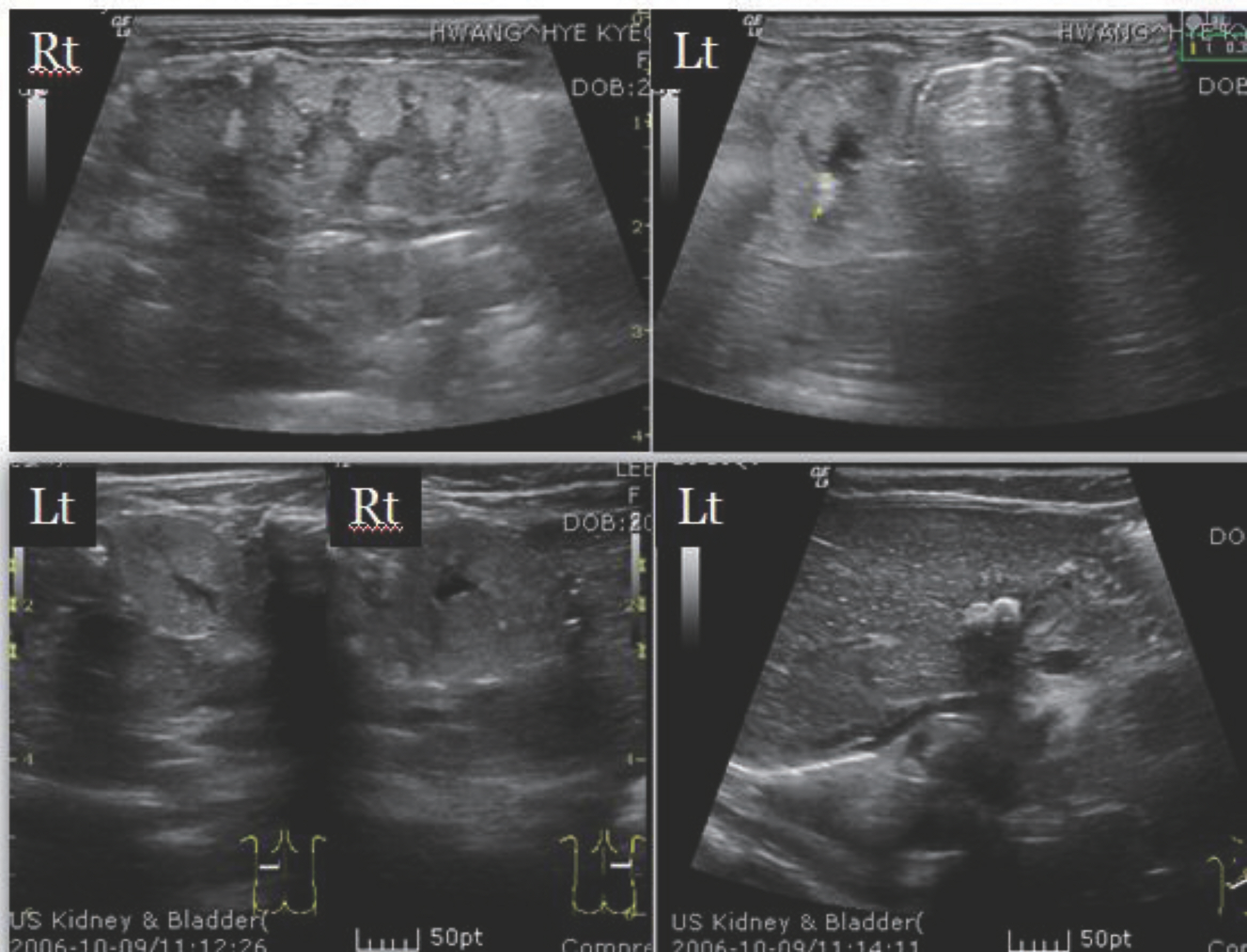
Risk Factors and Outcome of Nephrocalcinosis in Very Low Birth Weight Infants
- Affiliations
-
- 1Department of Pediatrics, Chonnam National University Hospital, Gwangju, Korea. essong@jnu.ac.kr
- KMID: 2072335
- DOI: http://doi.org/10.14734/kjp.2015.26.1.35
Abstract
- PURPOSE
The aim of this study was to determine the incidence, risk factors, and long-term outcome of nephrocalcinosis in very low birth weight (VLBW) infants.
METHODS
A retrospective chart review was performed in VLBW infants between 2006 and 2012 in the neonatal intensive care unit.
RESULTS
The incidence of nephrocalcinosis in VLBW infants was 10.2%. By univariate analysis, oligohydramnios and use of antenatal steroids were more frequent in the nephrocalcinosis group. In the nephrocalcinosis group, the gestational age and birth weight were lower and there were more number of female infants. Also, the initial blood pH, the lowest systolic blood pressure, and urine output on the first day of life were lower and bronchopulmonary dysplasia, sepsis, and urinary tract infection were more prevalent in the nephrocalcinosis group. The use of dexamethasone or ibuprofen and the lowest levels of phosphorus, protein and albumin were significantly lower in the nephrocalcinosis group. By binary logistic regression analysis, the use of antenatal steroids, female sex, 5-minute Apgar score, duration of oxygen therapy and total parenteral nutrition, and the lowest albumin level were found to be significant risk factors for nephrocalcinosis. Overall, the resolution rate was 64.1% and 88.6% within 12 months and 18 months, respectively.
CONCLUSION
The incidence of nephrocalcinosis in VLBW infants showed increasing trend. The risk factors of nephrocalcinosis were parameters for sick VLBW infants. Although the prognosis of nephrocalcinosis was relatively good, we should pay close attention to the development of complication.
MeSH Terms
-
Apgar Score
Birth Weight
Blood Pressure
Bronchopulmonary Dysplasia
Dexamethasone
Female
Gestational Age
Humans
Hydrogen-Ion Concentration
Ibuprofen
Incidence
Infant*
Infant, Newborn
Infant, Very Low Birth Weight*
Intensive Care, Neonatal
Logistic Models
Nephrocalcinosis*
Oligohydramnios
Oxygen
Parenteral Nutrition, Total
Phosphorus
Pregnancy
Prognosis
Retrospective Studies
Risk Factors*
Sepsis
Steroids
Urinary Tract Infections
Dexamethasone
Ibuprofen
Oxygen
Phosphorus
Steroids
Figure
Cited by 1 articles
-
Risk Factors for Nephrocalcinosis in Very Low Birth Weight Infants
Sang Eun Han, Teahyen Cha, Jinsup Kim, Ja Hye Kim, Chang Ryul Kim, Hyun Kyung Park, Hyun Ju Lee
Perinatology. 2019;30(1):14-19. doi: 10.14734/PN.2019.30.2.14.
- Full Text Links
- Actions
-
Cited
- CITED
-
- Close
- Share
- Similar articles
-
- Risk Factors for Nephrocalcinosis in Very Low Birth Weight Infants
- Risk Factors of Nephrocalcinosis in Very Low Birth Weight(VLBW) Infants
- Rehospitalization of Low-birth-weight Infants Who Were Discharged from NICU
- Furosemide-Induced Nephrocalcinosis in Very Low Birth Weight Infants
- Clinical Study of Prematurity and Low Birth Weight Infants


